Alzheimer’s disease
Antibody Makes Alzheimer’s Protein Detectable in Blood
Posted on by Dr. Francis Collins

Caption: The protein tau (green) aggregates abnormally in a brain cell (blue). Tau spills out of the cell and enters the bloodstream (red). Research shows that antibodies (blue) can capture tau in the blood that reflect its levels in the brain.
Credit: Sara Moser
Age can bring moments of forgetfulness. It can also bring concern that the forgetfulness might be a sign of early Alzheimer’s disease. For those who decide to have it checked out, doctors are likely to administer brief memory exams to assess the situation, and medical tests to search for causes of memory loss. Brain imaging and spinal taps can also help to look for signs of the disease. But an absolutely definitive diagnosis of Alzheimer’s disease is only possible today by examining a person’s brain postmortem. A need exists for a simple, less-invasive test to diagnose Alzheimer’s disease and similar neurodegenerative conditions in living people, perhaps even before memory loss becomes obvious.
One answer may lie in a protein called tau, which accumulates in abnormal tangles in the brains of people with Alzheimer’s disease and other “tauopathy” disorders. In recent years, researchers have been busy designing an antibody to target tau in hopes that this immunotherapy approach might slow or even reverse Alzheimer’s devastating symptoms, with promising early results in mice [1, 2]. Now, an NIH-funded research team that developed one such antibody have found it might also open the door to a simple blood test [3].
Snapshots of Life: Neurons in a New Light
Posted on by Dr. Francis Collins
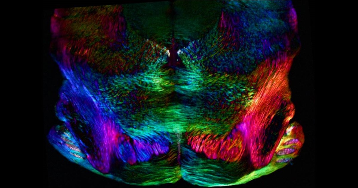
Credit: Michael Shribak, Marine Biological Laboratory, Woods Hole, MA
Birds do it, bees do it, and even educated fleas do it. No, not fall in love, as the late Ella Fitzgerald so famously sang. Birds and insects can see polarized light—that is, light waves transmitted in a single directional plane—in ways that provides them with a far more colorful and detailed view of the world than is possible with the human eye.
Still, thanks to innovations in microscope technology, scientists have been able to tap into the power of polarized light vision to explore the inner workings of many complex biological systems, including the brain. In this image, researchers used a recently developed polarized light microscope to trace the spatial orientation of neurons in a thin section of the mouse midbrain. Neurons that stretch horizontally appear green, while those oriented at a 45-degree angle are pinkish-red and those at 225 degrees are purplish-blue. What’s amazing is that these colors don’t involve staining or tagging the cells with fluorescent markers: the colors are generated strictly from the light interacting with the physical orientation of each neuron.
Aging Research: Plasma Protein Revitalizes the Brain
Posted on by Dr. Francis Collins
 For centuries, people have yearned for an elixir capable of restoring youth to their aging bodies and minds. It sounds like pure fantasy, but, in recent years, researchers have shown that the blood of young mice can exert a regenerative effect when transfused into older animals. Now, one of the NIH-funded teams that brought us those exciting findings has taken an early step toward extending them to humans.
In their latest work published in Nature, the researchers showed that blood plasma collected from the umbilical cords of newborn infants possesses some impressive rejuvenating effects [1]. When the human plasma was infused into the bloodstream of old mice, it produced marked improvements in learning and memory. Additional experiments traced many of those cognitive benefits to a specific protein called TIMP2—an unexpected discovery that could pave the way for the development of brain-boosting drugs to slow the effects of aging.
Exercise Releases Brain-Healthy Protein
Posted on by Dr. Francis Collins

We all know that exercise is important for a strong and healthy body. Less appreciated is that exercise seems also to be important for a strong and healthy mind, boosting memory and learning, while possibly delaying age-related cognitive decline [1]. How is this so? Researchers have assembled a growing body of evidence that suggests skeletal muscle cells secrete proteins and other factors into the blood during exercise that have a regenerative effect on the brain.
Now, an NIH-supported study has identified a new biochemical candidate to help explore the muscle-brain connection: a protein secreted by skeletal muscle cells called cathepsin B. The study found that levels of this protein rise in the blood of people who exercise regularly, in this case running on a treadmill. In mice, brain cells treated with the protein also exhibited molecular changes associated with the production of new neurons. Interestingly, the researchers found that the memory boost normally provided by exercise is diminished in mice unable to produce cathepsin B.
Creative Minds: A New Chemistry for Aging Research?
Posted on by Dr. Francis Collins

Tony Wyss-Coray / Credit: Stanford School of Medicine
Basic scientists have long studied aging by looking inside of cells. While this research has produced many important leads, they are now starting to look outside the cell for the wealth of biochemical clues contained in the bloodstream.
To introduce you to this exciting frontier in aging research, this blog highlighted a while back the work of Tony Wyss-Coray at Stanford School of Medicine, Palo Alto, CA. He and a colleague had just received a 2013 NIH Director’s Transformative Research Award to explore the effects of exercise on the brains of mice. Their work, in fact, produced one of Science Magazine’s Breakthrough Discoveries of 2014. Their team showed that by fusing the circulatory systems of old and young mice to create a shared blood supply, the young blood triggered new muscle and neural connections in the older mice, while also improving their memories.
As fascinating as this theoretical Fountain of Youth was, Wyss-Coray recognized a critical limitation. He had no way of knowing how factors secreted by the young mouse could actually cross the blood-brain barrier and rejuvenate neurons. To solve this unknown, Wyss-Coray recently received a 2015 NIH Director’s Pioneer Award to build a potentially game-changing tool to track the aging process in mice.
Alzheimer’s Disease: Tau Protein Predicts Early Memory Loss
Posted on by Dr. Francis Collins
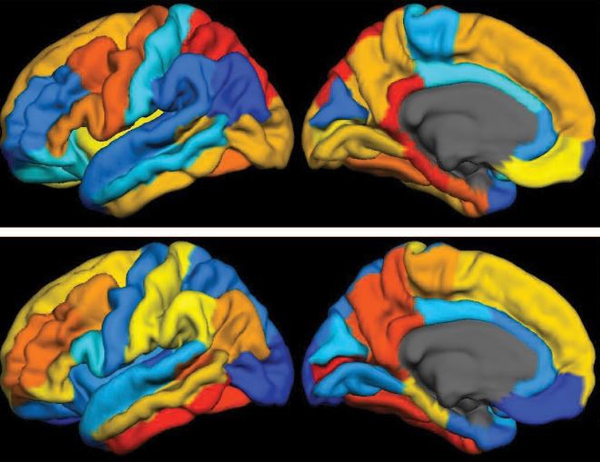
Caption: PET scan images show distribution of tau (top panel) and beta-amyloid (bottom panel) across a brain with early Alzheimer’s disease. Red indicates highest levels of protein binding, dark blue the lowest, yellows and oranges indicate moderate binding.
Credit: Brier et al., Sci Transl Med
In people with Alzheimer’s disease, changes in the brain begin many years before the first sign of memory problems. Those changes include the gradual accumulation of beta-amyloid peptides and tau proteins, which form plaques and tangles that are considered hallmarks of the disease. While amyloid plaques have received much attention as an early indicator of disease, until very recently there hadn’t been any way during life to measure the buildup of tau protein in the brain. As a result, much less is known about the timing and distribution of tau tangles and its relationship to memory loss.
Now, in a study published in Science Translational Medicine, an NIH-supported research team has produced some of the first maps showing where tau proteins build up in the brains of people with early Alzheimer’s disease [1]. The new findings suggest that while beta-amyloid remains a reliable early sign of Alzheimer’s disease, tau may be a more informative predictor of a person’s cognitive decline and potential response to treatment.
Creative Minds: Complex Solutions to Inflammation
Posted on by Dr. Francis Collins
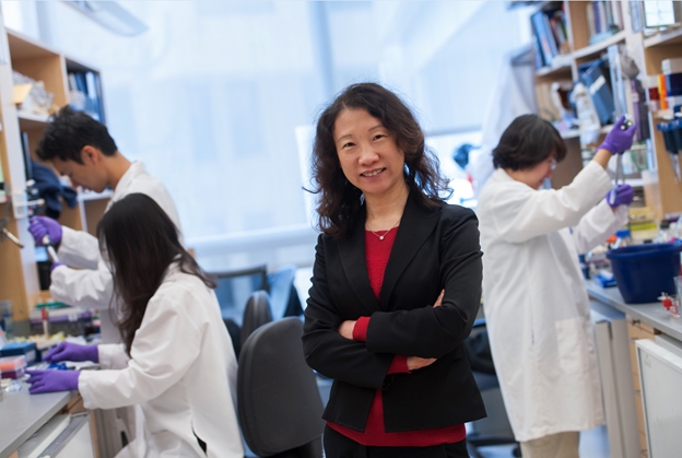
Hao Wu
For nearly 20 years, Hao Wu has studied innate immunity, our body’s first line of defense against infection. One of her research specialties is the challenging technique of X-ray crystallography, which she uses to capture the atomic structure of key molecules that drive an inflammatory response. But for this method to work, the proteins have to be coaxed to form regular crystals—and that has often proven to be prohibitively difficult. Wu, now at Boston Children’s Hospital and Harvard Medical School, can be relentless in her attempts to crystallize difficult molecular structures, and this quality has helped her make a number of important discoveries. Among them is the seminal finding that innate immune cells process and internalize signals to handle invading microbes much differently than previously thought.
Innate immune cells, which include macrophages and neutrophils, patrol the body non-specifically, keeping a look out for signs of anything unusual. Using protein receptors displayed on their surfaces, these cells can sense distinctive molecular patterns on microbes, prompting an immediate response at the site of infection.
Wu has shown that these cells form previously unknown protein complexes that mediate the immune response [1, 2]. She received an NIH Director’s 2015 Pioneer Award to help translate her expertise in the structural biology of these signaling complexes into the design of new kinds of anti-inflammatory treatments. This award helps exceptionally creative scientists to pioneer transformative approaches to major challenges in biomedical and behavioral research.
Got It Down Cold: Cryo-Electron Microscopy Named Method of the Year
Posted on by Dr. Francis Collins
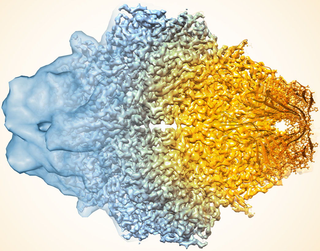
Credit: Veronica Falconieri, Subramaniam Lab, National Cancer Institute
In the quest to find faster, better ways of mapping the structure of proteins and other key biological molecules, a growing number of researchers are turning to an innovative method that pushes the idea of a freeze frame to a whole new level: cryo-electron microscopy (cryo-EM). The technique, which involves flash-freezing molecules in liquid nitrogen and bombarding them with electrons to capture their images with a special camera, has advanced dramatically since its inception thanks to the efforts of many creative minds. In fact, cryo-EM has improved so much that its mapping performance now rivals that of X-ray crystallography [1], the long-time workhorse of drug developers and structural biologists.
To get an idea of just how far cryo-EM has come over the last decade, take a look at the composite image above, which shows a bacterial enzyme (beta-galactosidase) bound to a drug-like molecule (phenylethyl beta-D-thiogalactopyranoside). To the left, you see a blob-like area generated by cryo-EM methods that would have been considered state-of-the-art just a few years ago. To the right, there’s an exquisitely detailed structure, which was produced at more than 10-times greater resolution using the latest advances in cryo-EM. In fact, today’s cryo-EM is so powerful that researchers can almost make out individual atoms! Very impressive, and just one of the many reasons why the journal Nature Methods recently named cryo-EM its “Method of the Year” for 2015 [2].
Creative Minds: Applying CRISPR Technology to Cancer Drug Resistance
Posted on by Dr. Francis Collins

Patrick Hsu
As a child, Patrick Hsu once settled a disagreement with his mother over antibacterial wipes by testing them in controlled experiments in the kitchen. When the family moved to Palo Alto, CA, instead of trying out for the football team or asking to borrow the family car like other high school kids might have done, Hsu went knocking on doors of scientists at Stanford University. He found his way into a neuroscience lab, where he gained experience with the fundamental tools of biology and a fascination for understanding how the brain works. But Hsu would soon become impatient with the tools that were available to ask some of the big questions he wanted to study.
As a Salk Helmsley Fellow and principal investigator at the Salk Institute for Biological Studies, La Jolla, CA, Hsu now works at the intersection of bioengineering, genomics, and neuroscience with a DNA editing tool called CRISPR/Cas9 that is revolutionizing the way scientists can ask and answer those big questions. (This blog has previously featured several examples of how this technology is revolutionizing biomedical research.) Hsu has received a 2015 NIH Director’s Early Independence award to adapt CRISPR/Cas9 technology so its use can be extended to that other critically important information-containing nucleic acid—RNA.Specifically, Hsu aims to develop ways to use this new tool to examine the role of a certain type of RNA in cancer drug resistance.
Creative Minds: Of Arsenic and Misfolded Proteins
Posted on by Dr. Francis Collins

John Hanna
Taking out the trash is a must in every household. Inside our cells, it’s also essential because if defective proteins are not properly disposed of, they can accumulate and make a mess of the cell’s inner workings, leading to health problems.
John Hanna, a physician-scientist at Brigham and Women’s Hospital, Boston, is on a quest to study the cell’s trash disposal system in greater detail. In particular, this 2014 NIH Director’s Early Independence awardee wants to learn more about how cells identify proteins that need to be discarded, how such proteins are steered towards the molecular garbage can, and how, when the process breaks down, neurodegenerative conditions, cancers, and other diseases can arise.
That’s a complex challenge, so Hanna will start by zeroing in on one particular component of cellular waste management—the component that clears out proteins damaged by arsenic. Although arsenic is notorious for being the poison of choice in countless true crime shows and mystery novels, this semi-metallic element is found naturally in soil, water, air, and some foods.
Previous Page Next Page