molecules
Got It Down Cold: Cryo-Electron Microscopy Named Method of the Year
Posted on by Dr. Francis Collins
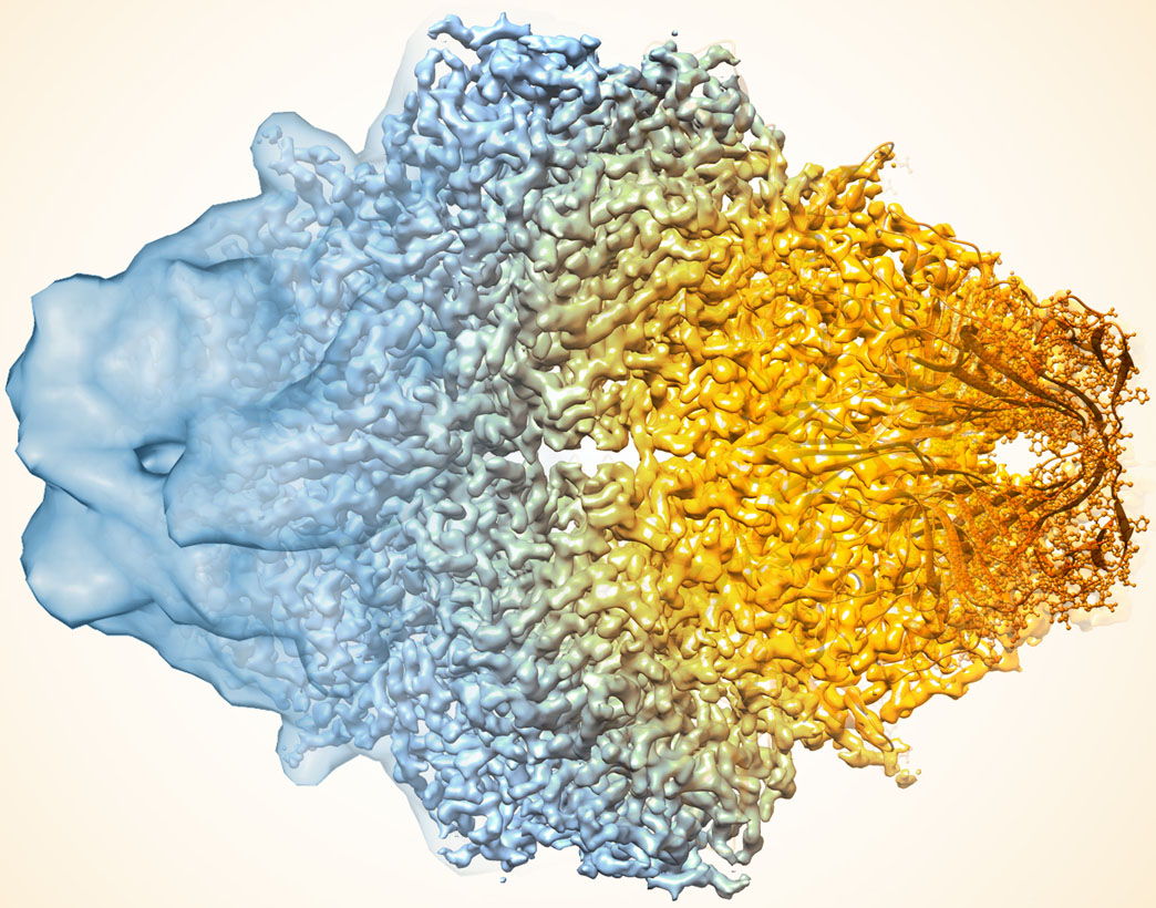
Credit: Veronica Falconieri, Subramaniam Lab, National Cancer Institute
In the quest to find faster, better ways of mapping the structure of proteins and other key biological molecules, a growing number of researchers are turning to an innovative method that pushes the idea of a freeze frame to a whole new level: cryo-electron microscopy (cryo-EM). The technique, which involves flash-freezing molecules in liquid nitrogen and bombarding them with electrons to capture their images with a special camera, has advanced dramatically since its inception thanks to the efforts of many creative minds. In fact, cryo-EM has improved so much that its mapping performance now rivals that of X-ray crystallography [1], the long-time workhorse of drug developers and structural biologists.
To get an idea of just how far cryo-EM has come over the last decade, take a look at the composite image above, which shows a bacterial enzyme (beta-galactosidase) bound to a drug-like molecule (phenylethyl beta-D-thiogalactopyranoside). To the left, you see a blob-like area generated by cryo-EM methods that would have been considered state-of-the-art just a few years ago. To the right, there’s an exquisitely detailed structure, which was produced at more than 10-times greater resolution using the latest advances in cryo-EM. In fact, today’s cryo-EM is so powerful that researchers can almost make out individual atoms! Very impressive, and just one of the many reasons why the journal Nature Methods recently named cryo-EM its “Method of the Year” for 2015 [2].