temporal lobe
The Brain Ripples Before We Remember
Posted on by Dr. Francis Collins

Throw a stone into a quiet pond, and you’ll see ripples expand across the water from the point where it went in. Now, neuroscientists have discovered that a different sort of ripple—an electrical ripple—spreads across the human brain when it strives to recall memories.
In memory games involving 14 very special volunteers, an NIH-funded team found that the split second before a person nailed the right answer, tiny ripples of electrical activity appeared in two specific areas of the brain [1]. If the volunteer recalled an answer incorrectly or didn’t answer at all, the ripples were much less likely to appear. While many questions remain, the findings suggest that the short, high-frequency electrical waves seen in these brain ripples may play an unexpectedly important role in our ability to remember.
The new study, published in Science, builds on brain recording data compiled over the last several years by neurosurgeon and researcher Kareem Zaghloul at NIH’s National Institute of Neurological Disorders and Stroke (NINDS). Zaghloul’s surgical team often temporarily places 10-to-20 arrays of tiny electrodes into the brains of a people with drug-resistant epilepsy. As I’ve highlighted recently, the brain mapping procedure aims to pinpoint the source of a patient’s epileptic seizures. But, with a patient’s permission, the procedure also presents an opportunity to learn more about how the brain works, with exceptional access to its circuits.
One such opportunity is to explore how the brain stores and recalls memories. To do this, the researchers show their patient volunteers hundreds of pairs of otherwise unrelated words, such as “pencil and bishop” or “orange and navy.” Later, they show them one of the words and test their memory to recall the right match. All the while, electrodes record the brain’s electrical activity.
Previously published studies by Zaghloul’s lab [2, 3] and many others have shown that memory involves the activation of a number of brain regions. That includes the medial temporal lobe, which is involved in forming and retrieving memories, and the prefrontal cortex, which helps in organizing memories in addition to its roles in “executive functions,” such as planning and setting goals. Those studies also have highlighted a role for the temporal association cortex, another portion of the temporal lobe involved in processing experiences and words.
In their data collected in patients with epilepsy, Zaghloul’s team’s earlier studies had uncovered some telltale patterns. For instance, when a person correctly recalled a word pair, the brain showed patterns of activity that looked quite similar to those present when he or she first learned to make a word association.
Alex Vaz, one of Zaghloul’s doctoral students, thought there might be more to the story. There was emerging evidence in rodents that brain ripples—short bursts of high frequency electrical activity—are involved in learning. There was also some evidence in people that such ripples might be important for solidifying memories during sleep. Vaz wondered whether they might find evidence of ripples as well in data gathered from people who were awake.
Vaz’s hunch was correct. The reanalysis revealed ripples of electricity in the medial temporal lobe and the temporal association cortex. When a person correctly recalled a word pair, those two brain areas rippled at the same time.
Further analysis showed that the ripples appeared in those two areas a few milliseconds before a volunteer remembered a word and gave a correct answer. Your brain is working on finding an answer before you are fully aware of it! Those ripples also appear to trigger brain waves that look similar to those observed in the association cortex when a person first learned a word pair.
The finding suggests that ripples in this part of the brain precede and may help to prompt the larger brain waves associated with replaying and calling to mind a particular memory. For example, hearing the words, “The Fab Four” may ripple into a full memory of a favorite Beatles album (yes! Sgt. Pepper’s Lonely Hearts Club Band) or, if you were lucky enough, a memorable concert back in the day (I never had that chance).
Zaghloul’s lab continues to study the details of these ripples to learn even more about how they may influence other neural signals and features involved in memory. So, the next time you throw a stone into a quiet pond and watch the ripples, perhaps it will trigger an electrical ripple in your brain to remember this blog and ruminate about this fascinating new discovery in neuroscience.
References:
[1] Coupled ripple oscillations between the medial temporal lobe and neocortex retrieve human memory. Vaz AP, Inati SK, Brunel N, Zaghloul KA. Science. 2019 Mar 1;363(6430):975-978.
[2] Cued Memory Retrieval Exhibits Reinstatement of High Gamma Power on a Faster Timescale in the Left Temporal Lobe and Prefrontal Cortex. Yaffe RB, Shaikhouni A, Arai J, Inati SK, Zaghloul KA. J Neurosci. 2017 Apr 26;37(17):4472-4480.
[3] Human Cortical Neurons in the Anterior Temporal Lobe Reinstate Spiking Activity during Verbal Memory Retrieval. Jang AI, Wittig JH Jr, Inati SK, Zaghloul KA. Curr Biol. 2017 Jun 5;27(11):1700-1705.e5.
Links:
Epilepsy Information Page (National Institute of Neurological Disorders and Stroke/NIH)
Brain Basics (NINDS)
Zaghloul Lab (NINDS)
NIH Support: National Institute of Neurological Disorders and Stroke; National Institute of General Medical Sciences
Alzheimer’s Disease: Tau Protein Predicts Early Memory Loss
Posted on by Dr. Francis Collins
Caption: PET scan images show distribution of tau (top panel) and beta-amyloid (bottom panel) across a brain with early Alzheimer’s disease. Red indicates highest levels of protein binding, dark blue the lowest, yellows and oranges indicate moderate binding.
Credit: Brier et al., Sci Transl Med
In people with Alzheimer’s disease, changes in the brain begin many years before the first sign of memory problems. Those changes include the gradual accumulation of beta-amyloid peptides and tau proteins, which form plaques and tangles that are considered hallmarks of the disease. While amyloid plaques have received much attention as an early indicator of disease, until very recently there hadn’t been any way during life to measure the buildup of tau protein in the brain. As a result, much less is known about the timing and distribution of tau tangles and its relationship to memory loss.
Now, in a study published in Science Translational Medicine, an NIH-supported research team has produced some of the first maps showing where tau proteins build up in the brains of people with early Alzheimer’s disease [1]. The new findings suggest that while beta-amyloid remains a reliable early sign of Alzheimer’s disease, tau may be a more informative predictor of a person’s cognitive decline and potential response to treatment.
Brain Imaging: Advance Aims for Epilepsy’s Hidden Hot Spots
Posted on by Dr. Francis Collins
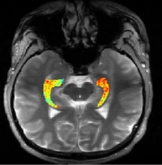
Credit: Reddy Lab, University of Pennsylvania
For many of the 65 million people around the world with epilepsy, modern medications are able to keep the seizures under control. When medications fail, as they do in about one-third of people with epilepsy, surgery to remove affected brain tissue without compromising function is a drastic step, but offers a potential cure. Unfortunately, not all drug-resistant patients are good candidates for such surgery for a simple reason: their brains appear normal on traditional MRI scans, making it impossible to locate precisely the source(s) of the seizures.
Now, in a small study published in Science Translational Medicine [1], NIH-funded researchers report progress towards helping such people. Using a new MRI method, called GluCEST, that detects concentrations of the nerve-signaling chemical glutamate in brain tissue [2], researchers successfully pinpointed seizure-causing areas of the brain in four of four volunteers with drug-resistant epilepsy and normal traditional MRI scans. While the findings are preliminary and must be confirmed by larger studies, researchers are hopeful that GluCEST, which takes about 30 minutes, may open the door to new ways of treating this type of epilepsy.