NIH Director’s Early Independence Award
Preventing Glaucoma Vision Loss with ‘Big Data’
Posted on by Dr. Francis Collins

Each morning, more than 2 million Americans start their rise-and-shine routine by remembering to take their eye drops. The drops treat their open-angle glaucoma, the most-common form of the disease, caused by obstructed drainage of fluid where the eye’s cornea and iris meet. The slow drainage increases fluid pressure at the front of the eye. Meanwhile, at the back of the eye, fluid pushes on the optic nerve, causing its bundled fibers to fray and leading to gradual loss of side vision.
For many, the eye drops help to lower intraocular pressure and prevent vision loss. But for others, the drops aren’t sufficient and their intraocular pressure remains high. Such people will need next-level care, possibly including eye surgery, to reopen the clogged drainage ducts and slow this disease that disproportionately affects older adults and African Americans over age 40.

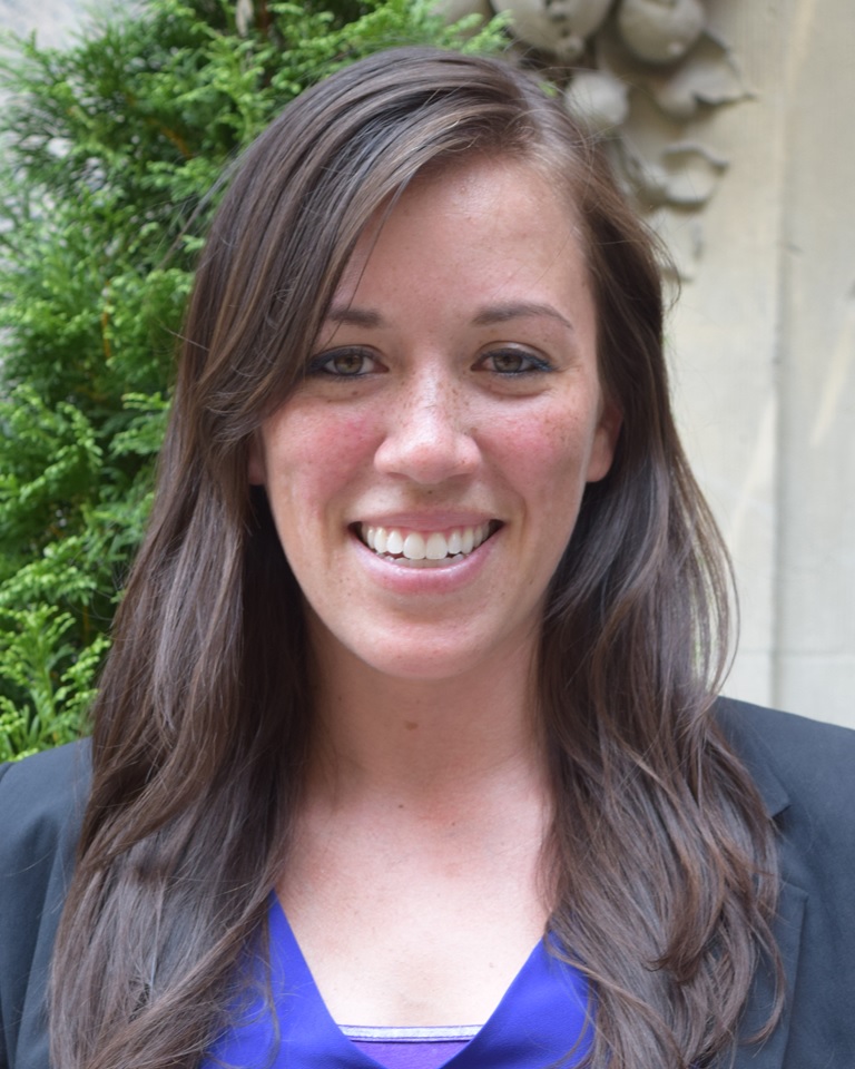
Credit: University of California San Diego
Sally Baxter, a physician-scientist with expertise in ophthalmology at the University of California, San Diego (UCSD), wants to learn how to predict who is at greatest risk for serious vision loss from open-angle and other forms of glaucoma. That way, they can receive more aggressive early care to protect their vision from this second-leading cause of blindness in the U.S..
To pursue this challenging research goal, Baxter has received a 2020 NIH Director’s Early Independence Award. Her research will build on the clinical observation that people with glaucoma frequently battle other chronic health problems, such as high blood pressure, diabetes, and heart disease. To learn more about how these and other chronic health conditions might influence glaucoma outcomes, Baxter has begun mining a rich source of data: electronic health records (EHRs).
In an earlier study of patients at UCSD, Baxter showed that EHR data helped to predict which people would need glaucoma surgery within the next six months [1]. The finding suggested that the EHR, especially information on a patient’s blood pressure and medications, could predict the risk for worsening glaucoma.
In her NIH-supported work, she’s already extended this earlier “Big Data” finding by analyzing data from more than 1,200 people with glaucoma who participate in NIH’s All of Us Research Program [2]. With consent from the participants, Baxter used their EHRs to train a computer to find telltale patterns within the data and then predict with 80 to 99 percent accuracy who would later require eye surgery.
The findings confirm that machine learning approaches and EHR data can indeed help in managing people with glaucoma. That’s true even when the EHR data don’t contain any information specific to a person’s eye health.
In fact, the work of Baxter and other groups have pointed to an especially important role for blood pressure in shaping glaucoma outcomes. Hoping to explore this lead further with the support of her Early Independence Award, Baxter also will enroll patients in a study to test whether blood-pressure monitoring smart watches can add important predictive information on glaucoma progression. By combining round-the-clock blood pressure data with EHR data, she hopes to predict glaucoma progression with even greater precision. She’s also exploring innovative ways to track whether people with glaucoma use their eye drops as prescribed, which is another important predictor of the risk of irreversible vision loss [3].
Glaucoma research continues to undergo great progress. This progress ranges from basic research to the development of new treatments and high-resolution imaging technologies to improve diagnostics. But Baxter’s quest to develop practical clinical tools hold great promise, too, and hopefully will help one day to protect the vision of millions of people with glaucoma around the world.
References:
[1] Machine learning-based predictive modeling of surgical intervention in glaucoma using systemic data from electronic health records. Baxter SL, Marks C, Kuo TT, Ohno-Machado L, Weinreb RN. Am J Ophthalmol. 2019 Dec; 208:30-40.
[2] Predictive analytics for glaucoma using data from the All of Us Research Program. Baxter SL, Saseendrakumar BR, Paul P, Kim J, Bonomi L, Kuo TT, Loperena R, Ratsimbazafy F, Boerwinkle E, Cicek M, Clark CR, Cohn E, Gebo K, Mayo K, Mockrin S, Schully SD, Ramirez A, Ohno-Machado L; All of Us Research Program Investigators. Am J Ophthalmol. 2021 Jul;227:74-86.
[3] Smart electronic eyedrop bottle for unobtrusive monitoring of glaucoma medication adherence. Aguilar-Rivera M, Erudaitius DT, Wu VM, Tantiongloc JC, Kang DY, Coleman TP, Baxter SL, Weinreb RN. Sensors (Basel). 2020 Apr 30;20(9):2570.
Links:
Glaucoma (National Eye Institute/NIH)
All of Us Research Program (NIH)
Video: Sally Baxter (All of Us Research Program)
Sally Baxter (University of California San Diego)
Baxter Project Information (NIH RePORTER)
NIH Director’s Early Independence Award (Common Fund)
NIH Support: Common Fund
Exploring the Universality of Human Song
Posted on by Dr. Francis Collins

It’s often said that music is a universal language. But is it really universal? Some argue that humans are just too culturally complex and their music is far too varied to expect any foundational similarity. Yet some NIH-funded researchers recently decided to take on the challenge, using the tools of computational social science to analyze recordings of human songs and other types of data gathered from more than 300 societies around the globe.
In a study published in the journal Science [1], the researchers conclude that music is indeed universal. Their analyses showed that all of the cultures studied used song in four similar behavioral contexts: dance, love, healing, and infant care. What’s more, no matter where in the world one goes, songs used in each of those ways were found to share certain musical features, including tone, pitch, and rhythm.
As exciting as the new findings may be for those who love music (like me), the implications may extend far beyond music itself. The work may help to shed new light on the complexities of the human brain, as well as inform efforts to enhance the role of music in improving human health. The healing power of music is a major focus of the NIH-supported Sound Health Initiative.
Samuel Mehr, a researcher at Harvard University, Cambridge, MA, led this latest study, funded in part by an NIH Director’s Early Independence Award. His multi-disciplinary team included anthropologists Manvir Singh, Harvard, and Luke Glowacki, Penn State University, State College; computational linguist Timothy O’Donnell, McGill University, Montreal, Canada; and political scientists Dean Knox, Princeton University, Princeton, NJ, and Christopher Lucas, Washington University, St. Louis.
In work published last year [2], Mehr’s team found that untrained listeners in 60 countries could on average discern the human behavior associated with culturally unfamiliar musical forms. These behaviors included dancing, soothing a baby, seeking to heal illness, or expressing love to another person.
In the latest study, the team took these initial insights and applied them more broadly to the universality of music. They started with the basic question: Do all human societies make music?
To find the answer, the team accessed Yale University’s Human Relations Area Files, an internationally recognized database for cultural anthropologists. This rich resource contains high-quality data for 319 mostly tribal societies across the planet, allowing the researchers to search archival information for mentions of music. Their search pulled up music tags for 309 societies. Digging deeper in other historical records not in the database, the team confirmed that the remaining six societies did indeed make music.
The researchers propose that these 319 societies provide a representative cross section of humanity. They thus conclude that it is statistically probable that music is in fact found in all human societies.
What exactly is so universal about music? To begin answering this complex question, the researchers tapped into more than a century of musicology to build a vast, multi-faceted database that they call the Natural History of Song (NHS).
Drawing from the NHS database, the researchers focused on nearly 5,000 vocally performed songs from 60 carefully selected human societies on all continents. By statistically analyzing those musical descriptions, they found that the behaviors associated with songs vary along three dimensions, which the researchers refer to as formality, arousal, and religiosity.
When the researchers mapped the four types of songs from their earlier study—love, dance, lullaby, and healing—onto these dimensions, they found that songs used in similar behavioral contexts around the world clustered together. For instance, across human societies, dance songs tend to appear in more formal contexts with large numbers of people. They also tend to be upbeat and energetic and don’t usually appear as part of religious ceremonies. In contrast, love songs tend to be more informal and less energetic.
Interestingly, the team also replicated its previous study in a citizen-science experiment with nearly 30,000 participants living in over 100 countries worldwide. They found again that listeners could tell what kinds of songs they were listening to, even when those songs came from faraway places. They went on to show that certain acoustic features of songs, like tempo, melody, and pitch, help to predict a song’s primary behavioral function across societies.
In many musical styles, melodies are composed of a fixed set of distinct tones organized around a tonal center (sometimes called the “tonic,” it’s the “do” in “do-re-mi”). For instance, the researchers explain, the tonal center of “Row Your Boat” is found in each “row” as well as the last “merrily,” and the final “dream.”
Their analyses show that songs with such basic tonal melodies are widespread and perhaps even universal. This suggests that tonality could be a means to delve even deeper into the natural history of world music and other associated behaviors, such as play, mourning, and fighting.
While some aspects of music may be universal, others are quite diverse. That’s particularly true within societies, where people may express different psychological states in song to capture their views of their culture. In fact, Mehr’s team found that the musical variation within a typical society is six times greater for that reason than the musical diversity across societies.
Following up on this work, Mehr’s team is now recruiting families with young infants for a study to understand how they respond to their varied collection of songs. Meanwhile, through the Sound Health Initiative, other research teams around the country are exploring many other ways in which listening to and creating music may influence and improve our health. As a scientist and amateur musician, I couldn’t be more excited to take part in this exceptional time of discovery at the intersection of health, neuroscience, and music.
References:
[1] Universality and diversity in human song. Mehr SA, Singh M, Knox D, Ketter DM, Pickens-Jones D, Atwood S, Lucas C, Jacoby N, Egner AA, Hopkins EJ, Howard RM, Hartshorne JK, Jennings MV, Simson J, Bainbridge CM, Pinker S, O’Donnell TJ, Krasnow MM, Glowacki L. Science. 2019 Nov 22;366(6468).
[2] Form and function in human song. Mehr SA, Singh M, York H, Glowacki L, Krasnow MM. Curr Biol. 2018 Feb 5;28(3):356-368.e5.
Links:
Sound Health Initiative (NIH)
Video: Music and the Mind—A Q & A with Renée Fleming & Francis Collins (YouTube)
The Music Lab (Harvard University, Cambridge, MA)
Samuel Mehr (Harvard)
NIH Director’s Early Independence Award (Common Fund)
NIH Support: Common Fund
What a Memory Looks Like
Posted on by Dr. Francis Collins

Your brain has the capacity to store a lifetime of memories, covering everything from the name of your first pet to your latest computer password. But what does a memory actually look like? Thanks to some very cool neuroscience, you are looking at one.
The physical manifestation of a memory, or engram, consists of clusters of brain cells active when a specific memory was formed. Your brain’s hippocampus plays an important role in storing and retrieving these memories. In this cross-section of a mouse hippocampus, imaged by the lab of NIH-supported neuroscientist Steve Ramirez, at Boston University, cells belonging to an engram are green, while blue indicates those not involved in forming the memory.
When a memory is recalled, the cells within an engram reactivate and turn on, to varying degrees, other neural circuits (e.g., sight, sound, smell, emotions) that were active when that memory was recorded. It’s not clear how these brain-wide connections are made. But it appears that engrams are the gatekeepers that mediate memory.
The story of this research dates back several years, when Ramirez helped develop a system that made it possible to image engrams by tagging cells in the mouse brain with fluorescent dyes. Using an innovative technology developed by other researchers, called optogenetics, Ramirez’s team then discovered it could shine light onto a collection of hippocampal neurons storing a specific memory and reactivate the sensation associated with the memory [1].
Ramirez has since gone on to show that, at least in mice, optogenetics can be used to trick the brain into creating a false memory [2]. From this work, he has also come to the interesting and somewhat troubling conclusion that the most accurate memories appear to be the ones that are never recalled. The reason: the mammalian brain edits—and slightly changes—memories whenever they are accessed.
All of the above suggested to Ramirez that, given its tremendous plasticity, the brain may possess the power to downplay a traumatic memory or to boost a pleasant recollection. Toward that end, Ramirez’s team is now using its mouse system to explore ways of suppressing one engram while enhancing another [3].
For Ramirez, though, the ultimate goal is to develop brain-wide maps that chart all of the neural networks involved in recording, storing, and retrieving memories. He recently was awarded an NIH Director’s Transformative Research Award to begin the process. Such maps will be invaluable in determining how stress affects memory, as well as what goes wrong in dementia and other devastating memory disorders.
References:
[1] Optogenetic stimulation of a hippocampal engram activates fear memory recall. Liu X, Ramirez S, Pang PT, Puryear CB, Govindarajan A, Deisseroth K, Tonegawa S. Nature. 2012 Mar 22;484(7394):381-385.
[2] Creating a false memory in the hippocampus. Ramirez S, Liu X, Lin PA, Suh J, Pignatelli M, Redondo RL, Ryan TJ, Tonegawa S. Science. 2013 Jul 26;341(6144):387-391.
[3] Artificially Enhancing and Suppressing Hippocampus-Mediated Memories. Chen BK, Murawski NJ, Cincotta C, McKissick O, Finkelstein A, Hamidi AB, Merfeld E, Doucette E, Grella SL, Shpokayte M, Zaki Y, Fortin A, Ramirez S. Curr Biol. 2019 Jun 3;29(11):1885-1894.
Links:
The Ramirez Group (Boston University, MA)
Ramirez Project Information (Common Fund/NIH)
NIH Director’s Early Independence Award (Common Fund)
NIH Director’s Transformative Research Award (Common Fund)
NIH Support: Common Fund
Creative Minds: Do Celebrity Endorsements Influence Teens’ Health?
Posted on by Dr. Francis Collins
Marie Bragg is a first-generation American, raised by a mother who immigrated to Florida from Trinidad. She watched her uncle in Florida cope effectively with type 2 diabetes, taking prescription drugs and following doctor-recommended dietary changes. But several of her Trinidadian relatives also had type 2 diabetes, and often sought to manage their diabetes by alternative means—through home remedies and spiritual practices.
This situation prompted Bragg to develop, at an early age, a strong interest in how approaches to health care may differ between cultures. But that wasn’t Bragg’s only interest—her other love was sports, having played on a high school soccer team that earned two state championships in Florida. That made her keenly aware of the sway that celebrity athletes, such as Michael Jordan and Serena Williams, could have on the public, particularly on young people. Today, Bragg combines both of her childhood interests—the influence of celebrities and the power of cultural narratives—in research that she is conducting as an Assistant Professor of Population Health at New York University Langone Medical Center and as a 2015 recipient of an NIH Director’s Early Independence Award.
Creative Minds: Applying CRISPR Technology to Cancer Drug Resistance
Posted on by Dr. Francis Collins

Patrick Hsu
As a child, Patrick Hsu once settled a disagreement with his mother over antibacterial wipes by testing them in controlled experiments in the kitchen. When the family moved to Palo Alto, CA, instead of trying out for the football team or asking to borrow the family car like other high school kids might have done, Hsu went knocking on doors of scientists at Stanford University. He found his way into a neuroscience lab, where he gained experience with the fundamental tools of biology and a fascination for understanding how the brain works. But Hsu would soon become impatient with the tools that were available to ask some of the big questions he wanted to study.
As a Salk Helmsley Fellow and principal investigator at the Salk Institute for Biological Studies, La Jolla, CA, Hsu now works at the intersection of bioengineering, genomics, and neuroscience with a DNA editing tool called CRISPR/Cas9 that is revolutionizing the way scientists can ask and answer those big questions. (This blog has previously featured several examples of how this technology is revolutionizing biomedical research.) Hsu has received a 2015 NIH Director’s Early Independence award to adapt CRISPR/Cas9 technology so its use can be extended to that other critically important information-containing nucleic acid—RNA.Specifically, Hsu aims to develop ways to use this new tool to examine the role of a certain type of RNA in cancer drug resistance.
Creative Minds: Fighting Cancer with Supercomputers
Posted on by Dr. Francis Collins
Amanda Randles
After graduating college with degrees in physics and computer science, Amanda Randles landed her dream first job. She joined IBM in 2005 to work on its Blue Gene Project, which had just unveiled the world’s fastest supercomputer. So fast, in fact, it’s said that a scientist with a calculator would have to work nonstop for 177,000 years to perform the operations that Blue Gene could complete in one second. As a member of the applications team, Randles was charged with writing new code to make the next model run even faster.
Randles left IBM in 2009 for graduate school, with the goal to apply her supercomputing expertise to biomedical research. She spent the next several years developing the necessary algorithms to produce a high-resolution 3D model of the human cardiovascular system, complete with realistic blood flow. Now, an assistant professor at Duke University, Durham, NC, and a 2014 NIH Director’s Early Independence awardee, Randles will build on her earlier work to attempt something even more challenging: simulating the movement of cancer cells through the circulation to predict where a tumor is most likely to spread. Randles hopes all of her late nights writing code will one day lead to software that helps doctors stage cancer more precisely and gives patients accurate personalized computer simulations that put an earlier, potentially life-saving bullseye on secondary tumors.
Creative Minds: Harnessing Technologies to Study Air Pollution’s Health Risks
Posted on by Dr. Francis Collins
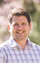
Perry Hystad
Credit: Hannah O’Leary, Oregon State University
After college, Perry Hystad took a trip to India and, while touring several large cities, noticed the vast clouds of exhaust from vehicles, smoke from factories, and soot from biomass-burning cook stoves. As he watched the rapid urban expansion all around him, Hystad remembers thinking: What effect does breathing such pollution day in and day out have upon these people’s health?
This question stuck with Hystad, and he soon developed a profound interest in environmental health. In 2013, Hystad completed his Ph.D. in his native Canada, studying the environmental risk factors for lung cancer [1, 2, 3]. Now, with the support of an NIH Director’s Early Independence Award, Hystad has launched his own lab at Oregon State University, Corvallis, to investigate further the health impacts of air pollution, which one recent analysis indicates may contribute to as many as several million deaths worldwide each year [4].
Creative Minds: Opening a Window on Alzheimer’s Before It Strikes
Posted on by Dr. Francis Collins

While attending college in her native Colombia, Yakeel T. Quiroz joined the Grupo de Neurociencias de Antioquia. This dedicated group of Colombian researchers, healthcare workers, and students has worked for many years with a large extended family in the northwestern district of Antioquia that is truly unique.
About half of the more than 5,000 family members inherit a gene mutation that predisposes them to what is known locally as “la bobera,” or “the foolishness,” a devastating form of early-onset Alzheimer’s disease. Those born with the mutation are cognitively healthy through their 20s, become forgetful in their 30s, and descend into full-blown Alzheimer’s disease by their mid-to-late 40s. Making matters worse, multiple family members sometimes are in different stages of dementia at the same time, including the caregiver attempting to hold the household together.
Quiroz, now a researcher at Massachusetts General Hospital, Boston, vowed never to forget these families. She hasn’t, working hard to understand early-onset Alzheimer’s disease and helping to establish the Forget Me Not Initiative to raise money for affected families. With an NIH Director’s Early Independence Award, Quiroz also recently launched her own lab to pursue an even broader scientific opportunity: discover subtle pre-symptomatic changes in the brain years before they give rise to detectable Alzheimer’s. What she learns will have application not only to detect and possibly treat early-onset Alzheimer’s in Colombia but also to understand the late-onset forms of the dementia that affect an estimated 35.6 million people worldwide.