Autism Spectrum Disorder
New Technology Opens Evolutionary Window into Brain Development
Posted on by Dr. Francis Collins

One of the great mysteries in biology is how we humans ended up with such large, complex brains. In search of clues, researchers have spent years studying the protein-coding genes activated during neurodevelopment. But some answers may also be hiding in non-coding regions of the human genome, where sequences called regulatory elements increase or decrease the activity of genes.
A fascinating example involves a type of regulatory element called a human accelerated region (HAR). Although “human” is part of this element’s name, it turns out that the genomes of all vertebrates—not just humans—contain the DNA segments now designated as HARs.
In most organisms, HARs show a relatively low rate of mutation, which means these regulatory elements have been highly conserved across species throughout evolutionary time [1]. The big exception is Homo sapiens, in which HARs have exhibited a much higher rate of mutations.
The accelerated rate of HARs mutations observed in humans suggest that, over the course of very long periods of time, these genomic changes might have provided our species with some sort of evolutionary advantage. What might that be? Many have speculated the advantage might involve the brain because HARs are often associated with genes involved in neurodevelopment. Now, in a paper published in the journal Neuron, an NIH-supported team confirms that’s indeed the case [2].
In the new work, researchers found that about half of the HARs in the human genome influence the activity, or expression, of protein-coding genes in neural cells and tissues during the brain’s development [3]. The researchers say their study—the most comprehensive to date of the 3,171 HARs in the human genome—firmly establishes that this type of regulatory element helps to drive patterns of neurodevelopmental gene activity specific to humans.
Yet to be determined is precisely how HARs affect the development of the human brain. The quest to uncover these details will no doubt shed new light on fundamental questions about the brain, its billions of neurons, and their trillions of interconnections. For example, why does human neural development span decades, longer than the life spans of most primates and other mammals? Answering such questions could also reveal new clues into a range of cognitive and behavioral disorders. In fact, early research has already made tentative links between HARs and neurodevelopmental conditions such as autism spectrum disorder and schizophrenia [3].
The latest work was led by Kelly Girskis, Andrew Stergachis, and Ellen DeGennaro, all of whom were in the lab of Christopher Walsh while working on the project. An NIH grantee, Walsh is director of the Allen Discovery Center for Brain Evolution at Boston Children’s Hospital and Harvard Medical School, which is supported by the Paul G. Allen Foundation Frontiers Group, and is an Investigator of the Howard Hughes Medical Institute.
Though HARs have been studied since 2006, one of the big challenges in systematically assessing them has been technological. The average length of a HAR is about 269 bases of DNA, but current technologies for assessing function can only easily analyze DNA molecules that span 150 bases or less.
Ryan Doan, who was then in the Walsh Lab, and his colleagues solved the problem by creating a new machine called CaptureMPRA. (MPRA is short for “massively parallel reporter assays.”) This technological advance cleverly barcodes HARs and, more importantly, makes it possible to analyze HARs up to about 500 bases in length.
Using CaptureMPRA technology in tandem with cell culture studies, researchers rolled up their sleeves and conducted comprehensive, full-sequence analyses of more than 3,000 HARs. In their initial studies, primarily in neural cells, they found nearly half of human HARs are active to drive gene expression in cell culture. Of those, 42 percent proved to have increased ability to enhance gene expression compared to their orthologues, or counterparts, in chimpanzees.
Next, the team integrated these data with an existing epigenetic dataset derived from developing human brain cells, as well as additional datasets generated from sorted brain cell types. They found that many HARs appeared to have the ability to increase the activity of protein-coding genes, while a smaller—but very significant—subset of the HARs appeared to be enhancing gene expression specifically in neural progenitor cells, which are responsible for making various neural cell types.
The data suggest that as the human HAR sequences mutated and diverged from other mammals, they increased their ability to enhance or sometimes suppress the activity of certain genes in neural cells. To illustrate this point, the researchers focused on two HARs that appear to interact specifically with a gene referred to as R17. This gene can have highly variable gene expression patterns not only in different human cell types, but also in cells from other vertebrates and non-vertebrates.
In the human cerebral cortex, the outermost part of the brain that’s responsible for complex behaviors, R17 is expressed only in neural progenitor cells and only at specific time points. The researchers found that R17 slows the progression of neural progenitor cells through the cell cycle. That might seem strange, given the billions of neurons that need to be made in the cortex. But it’s consistent with the biology. In the human, it takes more than 130 days for the cortex to complete development, compared to about seven days in the mouse.
Clearly, to learn more about how the human brain evolved, researchers will need to look for clues in many parts of the genome at once, including its non-coding regions. To help researchers navigate this challenging terrain, the Walsh team has created an online resource displaying their comprehensive HAR data. It will appear soon, under the name HAR Hub, on the University of California Santa Cruz Genome Browser.
References:
[1] An RNA gene expressed during cortical development evolved rapidly in humans. Pollard KS, Salama SR, Lambert N, Lambot MA, Coppens S, Pedersen JS, Katzman S, King B, Onodera C, Siepel A, Kern AD, Dehay C, Igel H, Ares M Jr, Vanderhaeghen P, Haussler D. Nature. 2006 Sep 14;443(7108):167-72.
[2] Rewiring of human neurodevelopmental gene regulatory programs by human accelerated regions. Girskis KM, Stergachis AB, DeGennaro EM, Doan RN, Qian X, Johnson MB, Wang PP, Sejourne GM, Nagy MA, Pollina EA, Sousa AMM, Shin T, Kenny CJ, Scotellaro JL, Debo BM, Gonzalez DM, Rento LM, Yeh RC, Song JHT, Beaudin M, Fan J, Kharchenko PV, Sestan N, Greenberg ME, Walsh CA. Neuron. 2021 Aug 25:S0896-6273(21)00580-8.
[3] Mutations in human accelerated regions disrupt cognition and social behavior. Doan RN, Bae BI, Cubelos B, Chang C, Hossain AA, Al-Saad S, Mukaddes NM, Oner O, Al-Saffar M, Balkhy S, Gascon GG; Homozygosity Mapping Consortium for Autism, Nieto M, Walsh CA. Cell. 2016 Oct 6;167(2):341-354.
Links:
Christopher Walsh Laboratory (Boston Children’s Hospital and Harvard Medical School)
The Paul G. Allen Foundation Frontiers Group (Seattle)
NIH Support: National Institute of Neurological Disorders and Stroke; National Institute of Mental Health; National Institute of General Medical Sciences; National Cancer Institute
The Amazing Brain: Tracking Molecular Events with Calling Cards
Posted on by Dr. Francis Collins
In days mostly gone by, it was fashionable in some circles for people to hand out calling cards to mark their arrival at special social events. This genteel human tradition is now being adapted to the lab to allow certain benign viruses to issue their own high-tech calling cards and mark their arrival at precise locations in the genome. These special locations show where there’s activity involving transcription factors, specialized proteins that switch genes on and off and help determine cell fate.
The idea is that myriad, well-placed calling cards can track brain development over time in mice and detect changes in transcription factor activity associated with certain neuropsychiatric disorders. This colorful image, which won first place in this year’s Show Us Your BRAINs! Photo and Video contest, provides a striking display of these calling cards in action in living brain tissue.
The image comes from Allen Yen, a PhD candidate in the lab of Joseph Dougherty, collaborating with the nearby lab of Rob Mitra. Both labs are located in the Washington University School of Medicine, St. Louis.
Yen and colleagues zoomed in on this section of mouse brain tissue under a microscope to capture dozens of detailed images that they then stitched together to create this high-resolution overview. The image shows neural cells (red) and cell nuclei (blue). But focus in on the neural cells (green) concentrated in the brain’s outer cortex (top) and hippocampus (two lobes in the upper center). They’ve been labelled with calling cards that were dropped off by adeno-associated virus [1].
Once dropped off, a calling card doesn’t bear a pretentious name or title. Rather, the calling card, is a small mobile snippet of DNA called a transposon. It gets dropped off with the other essential component of the technology: a specialized enzyme called a transposase, which the researchers fuse to one of many specific transcription factors of interest.
Each time one of these transcription factors of interest binds DNA to help turn a gene on or off, the attached transposase “grabs” a transposon calling card and inserts it into the genome. As a result, it leaves behind a permanent record of the interaction.
What’s also nice is the calling cards are programmed to give away their general locations. That’s because they encode a fluorescent marker (in this image, it’s a green fluorescent protein). In fact, Yen and colleagues could look under a microscope and tell from all the green that their calling card technology was in place and working as intended.
The final step, though, was to find out precisely where in the genome those calling cards had been left. For this, the researchers used next-generation sequencing to produce a cumulative history and map of each and every calling card dropped off in the genome.
These comprehensive maps allow them to identify important DNA-protein binding events well after the fact. This innovative technology also enables scientists to attribute past molecular interactions with observable developmental outcomes in a way that isn’t otherwise possible.
While the Mitra and Dougherty labs continue to improve upon this technology, it’s already readily adaptable to answer many important questions about the brain and brain disorders. In fact, Yen is now applying the technology to study neurodevelopment in mouse models of neuropsychiatric disorders, specifically autism spectrum disorder (ASD) [2]. This calling card technology also is available for any lab to deploy for studying a transcription factor of interest.
This research is supported by the Brain Research through Advancing Innovative Neurotechnologies® (BRAIN) Initiative. One of the major goals of BRAIN Initiative is to accelerate the development and application of innovative technologies to gain new understanding of the brain. This award-winning image is certainly a prime example of striving to meet this goal. I’ll look forward to what these calling cards will tell us in the future about ASD and other important neurodevelopmental conditions affecting the brain.
References:
[1] A viral toolkit for recording transcription factor-DNA interactions in live mouse tissues. Cammack AJ, Moudgil A, Chen J, Vasek MJ, Shabsovich M, McCullough K, Yen A, Lagunas T, Maloney SE, He J, Chen X, Hooda M, Wilkinson MN, Miller TM, Mitra RD, Dougherty JD. Proc Natl Acad Sci U S A. 2020 May 5;117(18):10003-10014.
[2] A MYT1L Syndrome mouse model recapitulates patient phenotypes and reveals altered brain development due to disrupted neuronal maturation. Jiayang Chen, Mary E. Lambo, Xia Ge, Joshua T. Dearborn, Yating Liu, Katherine B. McCullough, Raylynn G. Swift, Dora R. Tabachnick, Lucy Tian, Kevin Noguchi, Joel R. Garbow, John N. Constantino. bioRxiv. May 27, 2021.
Links:
Brain Research through Advancing Innovative Neurotechnologies® (BRAIN) Initiative (NIH)
Autism Spectrum Disorder (National Institute of Mental Health/NIH)
Dougherty Lab (Washington University School of Medicine, St. Louis)
Mitra Lab (Washington University School of Medicine)
Show Us Your BRAINs! Photo and Video Contest (BRAIN Initiative/NIH)
NIH Support: National Institute of Neurological Disorders and Stroke; National Institute of Mental Health; National Center for Advancing Translational Sciences; National Human Genome Research Institute; National Institute of General Medical Sciences
Largest-Ever Genetic Study of Autism Yields New Insights
Posted on by Dr. Francis Collins

Anyone who’s spent time with people affected by autism spectrum disorder (ASD) can tell you that it’s a very complex puzzle. The wide variability seen among individuals with this group of developmental brain disorders, which can disrupt communication, behavior control, and social skills, has also posed a huge challenge for researchers trying to identify underlying genetic and environmental factors. So, it’s no surprise that there’s been considerable interest in the recent findings of the largest-ever genetic study of ASD.
In a landmark study that analyzed the DNA of more than 35,000 people from around the world, the NIH-funded international Autism Sequencing Consortium (ASC) identified variants in 102 genes associated with increased risk of developing ASD, up from 65 identified previously. Of the 102 genes, 60 had not been previously linked to ASD and 53 appeared to be primarily connected to ASD as opposed to other types of intellectual disability or developmental delay. It is expected that this newfound genetic knowledge will serve to improve understanding of the complex biological mechanisms involved in ASD, ultimately paving the way for new approaches to diagnosis and treatment.
The study reported in the journal Cell was led by Joseph Buxbaum, Icahn School of Medicine at Mount Sinai, New York; Stephan Sanders, University of California, San Francisco; Kathryn Roeder, Carnegie Mellon University, Pittsburgh, PA; and Mark Daly, Massachusetts General Hospital, Boston, MA and the Broad Institute of MIT and Harvard, Cambridge, MA. These researchers and their teams faced what might seem like a rather daunting task.
While common genetic variants collectively are known to contribute substantially to ASD, rare variants have been recognized individually as more major contributors to a person’s risk of developing ASD. The challenge was how to find such rare variants—whether inherited or newly arising.
To do so, the researchers needed to analyze a enormous amount of DNA data. Fortunately, they and their ASC colleagues already had assembled a vast trove of data. Over the last decade, the ASC had collected DNA samples with full consent from thousands of people with and without ASD, including unaffected siblings and parents. All were aggregated with other studies, and, at the time of this investigation, they had gathered 35,584 unique samples. Those included more than 21,000 family-based samples and almost 12,000 samples from people diagnosed with ASD.
In search of rare genetic alterations, they sequenced whole exomes, the approximately 1.5 percent of the genome that codes for proteins. Their search produced a list of 102 ASD-associated genes, including 30 that had never been implicated in any developmental brain disorder previously.
But that was just the beginning. Next, the ASC team dug deeper into this list. The researchers knew from previous work that up to half of people with ASD also have an intellectual disability or developmental delay. Many of the associated genes overlap, meaning they play roles in both outcomes. So, in one set of analyses, the team compared the list to the results of another genetic study of people diagnosed with developmental delays, including problems with learning or gross motor skills such as delayed walking.
The detailed comparison allowed them to discern genes that are more associated with features of ASD, as opposed to those that are more specific to these developmental delays. It turns out that 49 of the 102 autism-associated genes were altered more often in people with developmental delay than in those diagnosed with ASD. The other 53 were altered more often in ASD, suggesting that they may be more closely linked to this condition’s unique features.
Further study also showed that people who carried alterations in genes found predominantly in ASD also had better intellectual function. They also were more likely to have learned to walk without a developmental delay.
The 102 new genes fell primarily into one of two categories. Many play a role in the brain’s neural connections. The rest are involved primarily in switching other genes on and off in brain development. Interestingly, they are expressed both in excitatory neurons, which are active in sending signals in the brain, and in inhibitory neurons that squelch such activity. Many of these genes are also commonly expressed in the brain’s cerebral cortex, the outermost part of the brain that is responsible for many complex behaviors.
Overall, these findings underscore that ASD truly does exist on a spectrum. Indeed, there are many molecular paths to this disorder. The ASC researchers continue to collect samples, so we can expect this list of 102 genes will continue to expand in the future.
With these gene discoveries in hand, the researchers will now also turn their attention to unravelling additional details about how these genes function in the brain. The hope is that this growing list of genes will converge on a smaller number of important molecular pathways, pointing the way to new and more precise ways of treating ASD in all its complexity.
Reference:
[1] Large-scale exome sequencing study implicates both developmental and functional changes in the neurobiology of autism. Satterstrom FK, Kosmicki JA, Wang J, Breen MS, De Rubeis S, An JY, Peng M, Collins R, Grove J, Klei L, Stevens C, Reichert J, Mulhern MS, Artomov M, Gerges S, Sheppard B, Xu X, Bhaduri A, Norman U, Brand H, Schwartz G, Nguyen R, Guerrero EE, Dias C; Autism Sequencing Consortium; iPSYCH-Broad Consortium, Betancur C, Cook EH, Gallagher L, Gill M, Sutcliffe JS, Thurm A, Zwick ME, Børglum AD, State MW, Cicek AE, Talkowski ME, Cutler DJ, Devlin B, Sanders SJ, Roeder K, Daly MJ, Buxbaum JD.Cell. 2020 Jan 23. {Epub ahead of print]
Links:
Autism Spectrum Disorder (NIH/National Institute of Mental Health)
Joseph Buxbaum (Icahn School of Medicine at Mount Sinai, New York)
Sanders Lab (University of California, San Francisco)
Kathryn Roeder (Carnegie Mellon University, Pittsburgh, PA)
Mark Daly (Broad Institute of MIT and Harvard, Cambridge, MA)
Autism Sequencing Consortium (Emory University, Atlanta)
NIH Support: National Institute Mental Health; National Human Genome Research Institute
Could A Gut-Brain Connection Help Explain Autism?
Posted on by Dr. Francis Collins

You might think nutrient-sensing cells in the human gastrointestinal (GI) tract would have no connection whatsoever to autism spectrum disorder (ASD). But if Diego Bohórquez’s “big idea” is correct, these GI cells, called neuropods, could one day help to provide a direct link into understanding and treating some aspects of autism and other brain disorders.
Bohórquez, a researcher at Duke University, Durham, NC, recently discovered that cells in the intestine, previously known for their hormone-releasing ability, form extensions similar to neurons. He also found that those extensions connect to nerve fibers in the gut, which relay signals to the vagus nerve and onward to the brain. In fact, he found that those signals reach the brain in milliseconds [1].
Bohórquez has dedicated his lab to studying this direct, high-speed hookup between gut and brain and its impact on nutrient sensing, eating, and other essential behaviors. Now, with support from a 2019 NIH Director’s New Innovator Award, he will also explore the potential for treating autism and other brain disorders with drugs that act on the gut.
Bohórquez became interested in autism and its possible link to the gut-brain connection after a chance encounter with Geraldine Dawson, director of the Duke Center for Autism and Brain Development. Dawson mentioned that autism typically affects multiple organ systems.
With further reading, he discovered that kids with autism frequently cope with GI issues, including bowel inflammation, abdominal pain, constipation, and/or diarrhea [2]. They often also show unusual food-related behaviors, such as being extremely picky eaters. But his curiosity was especially piqued by evidence that certain gut microbes can influence abnormal behaviors in mice that model autism.
With his New Innovator Award, Bohórquez will study neuropods and the gut-brain connection in a mouse model of autism. Using the tools of optogenetics, which make it possible to activate cells with light, he’ll also see whether autism-like symptoms in mice can be altered or alleviated by controlling neuropods in the gut. Those symptoms include anxiety, repetitive behaviors, and lack of interest in interacting with other mice. He’ll also explore changes in the animals’ eating habits.
In another line of study, he will take advantage of intestinal tissue samples collected from people with autism. He’ll use those tissues to grow and then examine miniature intestinal “organoids,” looking for possible evidence that those from people with autism are different from others.
For the millions of people now living with autism, no truly effective drug therapies are available to help to manage the condition and its many behavioral and bodily symptoms. Bohórquez hopes one day to change that with drugs that act safely on the gut. In the meantime, he and his fellow “GASTRONAUTS” look forward to making some important and fascinating discoveries in the relatively uncharted territory where the gut meets the brain.
References:
[1] A gut-brain neural circuit for nutrient sensory transduction. Kaelberer MM, Buchanan KL, Klein ME, Barth BB, Montoya MM, Shen X, Bohórquez DV. Science. 2018 Sep 21;361(6408).
[2] Association of maternal report of infant and toddler gastrointestinal symptoms with autism: evidence from a prospective birth cohort. Bresnahan M, Hornig M, Schultz AF, Gunnes N, Hirtz D, Lie KK, Magnus P, Reichborn-Kjennerud T, Roth C, Schjølberg S, Stoltenberg C, Surén P, Susser E, Lipkin WI. JAMA Psychiatry. 2015 May;72(5):466-474.
Links:
Autism Spectrum Disorder (National Institute of Mental Health/NIH)
Bohórquez Lab (Duke University, Durham, NC)
Bohórquez Project Information (NIH RePORTER)
NIH Director’s New Innovator Award (Common Fund)
NIH Support: Common Fund; National Institute of Mental Health
A Neuronal Light Show
Posted on by Dr. Francis Collins

These colorful lights might look like a video vignette from one of the spectacular evening light shows taking place this holiday season. But they actually aren’t. These lights are illuminating the way to a much fuller understanding of the mammalian brain.
The video features a new research method called BARseq (Barcoded Anatomy Resolved by Sequencing). Created by a team of NIH-funded researchers led by Anthony Zador, Cold Spring Harbor Laboratory, NY, BARseq enables scientists to map in a matter of weeks the location of thousands of neurons in the mouse brain with greater precision than has ever been possible before.
How does it work? With BARseq, researchers generate uniquely identifying RNA barcodes and then tag one to each individual neuron within brain tissue. As reported recently in the journal Cell, those barcodes allow them to keep track of the location of an individual cell amid millions of neurons [1]. This also enables researchers to map the tangled paths of individual neurons from one region of the mouse brain to the next.
The video shows how the researchers read the barcodes. Each twinkling light is a barcoded neuron within a thin slice of mouse brain tissue. The changing colors from frame to frame correspond to one of the four letters, or chemical bases, in RNA (A=purple, G=blue, U=yellow, and C=white). A neuron that flashes blue, purple, yellow, white is tagged with a barcode that reads GAUC, while yellow, white, white, white is UCCC.
By sequencing and reading the barcodes to distinguish among seemingly identical cells, the researchers mapped the connections of more than 3,500 neurons in a mouse’s auditory cortex, a part of the brain involved in hearing. In fact, they report they’re now able to map tens of thousands of individual neurons in a mouse in a matter of weeks.
What makes BARseq even better than the team’s previous mapping approach, called MAPseq, is its ability to read the barcodes at their original location in the brain tissue [2]. As a result, they can produce maps with much finer resolution. It’s also possible to maintain other important information about each mapped neuron’s identity and function, including the expression of its genes.
Zador reports that they’re continuing to use BARseq to produce maps of other essential areas of the mouse brain with more detail than had previously been possible. Ultimately, these maps will provide a firm foundation for better understanding of human thought, consciousness, and decision-making, along with how such mental processes get altered in conditions such as autism spectrum disorder, schizophrenia, and depression.
Here’s wishing everyone a safe and happy holiday season. It’s been a fantastic year in science, and I look forward to bringing you more cool NIH-supported research in 2020!
References:
[1] High-Throughput Mapping of Long-Range Neuronal Projection Using In Situ Sequencing. Chen X, Sun YC, Zhan H, Kebschull JM, Fischer S, Matho K, Huang ZJ, Gillis J, Zador AM. Cell. 2019 Oct 17;179(3):772-786.e19.
[2] High-Throughput Mapping of Single-Neuron Projections by Sequencing of Barcoded RNA. Kebschull JM, Garcia da Silva P, Reid AP, Peikon ID, Albeanu DF, Zador AM. Neuron. 2016 Sep 7;91(5):975-987.
Links:
Brain Research through Advancing Innovative Neurotechnologies® (BRAIN) Initiative (NIH)
Zador Lab (Cold Spring Harbor Laboratory, Cold Spring Harbor, NY)
NIH Support: National Institute of Neurological Disorders and Stroke; National Institute on Drug Abuse; National Cancer Institute
Study Shows Genes Unique to Humans Tied to Bigger Brains
Posted on by Dr. Francis Collins
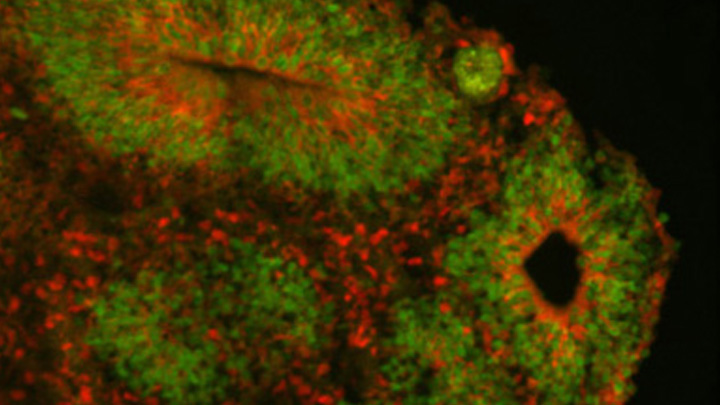
Caption: Cortical organoid, showing radial glial stem cells (green) and cortical neurons (red).
Credit: Sofie Salama, University of California, Santa Cruz
In seeking the biological answer to the question of what it means to be human, the brain’s cerebral cortex is a good place to start. This densely folded, outer layer of grey matter, which is vastly larger in Homo sapiens than in other primates, plays an essential role in human consciousness, language, and reasoning.
Now, an NIH-funded team has pinpointed a key set of genes—found only in humans—that may help explain why our species possesses such a large cerebral cortex. Experimental evidence shows these genes prolong the development of stem cells that generate neurons in the cerebral cortex, which in turn enables the human brain to produce more mature cortical neurons and, thus, build a bigger cerebral cortex than our fellow primates.
That sounds like a great advantage for humans! But there’s a downside. Researchers found the same genomic changes that facilitated the expansion of the human cortex may also render our species more susceptible to certain rare neurodevelopmental disorders.
Wearable Scanner Tracks Brain Activity While Body Moves
Posted on by Dr. Francis Collins
Credit: Wellcome Centre for Human Neuroimaging, University College London.
In recent years, researchers fueled by the BRAIN Initiative and many other NIH-supported efforts have made remarkable progress in mapping the human brain in all its amazing complexity. Now, a powerful new imaging technology promises to further transform our understanding [1]. This wearable scanner, for the first time, enables researchers to track neural activity in people in real-time as they do ordinary things—be it drinking tea, typing on a keyboard, talking to a friend, or even playing paddle ball.
This new so-called magnetoencephalography (MEG) brain scanner, which looks like a futuristic cross between a helmet and a hockey mask, is equipped with specialized “quantum” sensors. When placed directly on the scalp surface, these new MEG scanners can detect weak magnetic fields generated by electrical activity in the brain. While current brain scanners weigh in at nearly 1,000 pounds and require people to come to a special facility and remain absolutely still, the new system weighs less than 2 pounds and is capable of generating 3D images even when a person is making motions.
Studies of Dogs, Mice, and People Provide Clues to OCD
Posted on by Dr. Francis Collins

Thinkstock/wildpixel
Chances are you know someone with obsessive-compulsive disorder (OCD). It’s estimated that more than 2 million Americans struggle with this mental health condition, characterized by unwanted recurring thoughts and/or repetitive behaviors, such as excessive hand washing or constant counting of objects. While we know that OCD tends to run in families, it’s been frustratingly difficult to identify specific genes that influence OCD risk.
Now, an international research team, partly funded by NIH, has made progress thanks to an innovative genomic approach involving dogs, mice, and people. The strategy allowed them to uncover four genes involved in OCD that turn out to play a role in synapses, where nerve impulses are transmitted between neurons in the brain. While more research is needed to confirm the findings and better understand the molecular mechanisms of OCD, these findings offer important new leads that could point the way to more effective treatments.
How Kids See the World Depends a Lot on Genetics
Posted on by Dr. Francis Collins
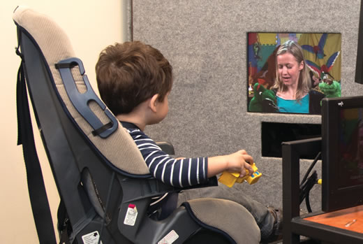
Caption: Child watches video while researchers track his eye movements.
Credit: Washington University School of Medicine, St. Louis
From the time we are born, most of us humans closely watch the world around us, paying special attention to people’s faces and expressions. Now, for the first time, an NIH-funded team has shown that the ways in which children look at faces and many other things are strongly influenced by the genes they’ve inherited from their parents.
The findings come from experiments that tracked the eye movements of toddlers watching videos of other kids or adult caregivers. The experiments showed that identical twins—who share the same genes and the same home environment—spend almost precisely the same proportion of time looking at faces, even when watching different videos. And when identical twins watched the same video, they tended to look at the same thing at almost exactly the same time! In contrast, fraternal twins—who shared the same home environment, but, on average, shared just half of their genes—had patterns of eye movement that were far less similar.
Interestingly, the researchers also found that the visual behaviors most affected in children with autism spectrum disorder (ASD)—attention to another person’s eyes and mouth—were those that also appeared to be the most heavily influenced by genetics. The discovery makes an important connection between two well-known features of ASD: a strong hereditary component and poor eye contact with other people.
Autism Spectrum Disorder: Progress Toward Earlier Diagnosis
Posted on by Dr. Francis Collins

Stockbyte
Research shows that the roots of autism spectrum disorder (ASD) generally start early—most likely in the womb. That’s one more reason, on top of a large number of epidemiological studies, why current claims about the role of vaccines in causing autism can’t be right. But how early is ASD detectable? It’s a critical question, since early intervention has been shown to help limit the effects of autism. The problem is there’s currently no reliable way to detect ASD until around 18–24 months, when the social deficits and repetitive behaviors associated with the condition begin to appear.
Several months ago, an NIH-funded team offered promising evidence that it may be possible to detect ASD in high-risk 1-year-olds by shifting attention from how kids act to how their brains have grown [1]. Now, new evidence from that same team suggests that neurological signs of ASD might be detectable even earlier.
Next Page