mental health
Changes in Normal Brain Connections Linked to Eating Disorders
Posted on by Lawrence Tabak, D.D.S., Ph.D.

Anyone who has ever had a bad habit knows how vexingly difficult breaking it can be. The reason is the repeated action, initially linked to some type of real or perceived reward, over time changes the way our very brains are wired to work. The bad habit becomes automatic, even when the action does us harm or we no longer wish to do it.
Now an intriguing new study shows that the same bundled nerve fibers, or brain circuits, involved in habit formation also can go awry in people with eating disorders. The findings may help to explain why eating disorders are so often resistant to will power alone. They also may help to point the way to improved approaches to treating eating disorders, suggesting strategies that adjust the actual brain circuitry in helpful ways.
These latest findings, published in the journal Science Translational Medicine, come from the NIH-supported Casey Halpern, University of Pennsylvania’s Perelman School of Medicine, Philadelphia, and Cara Bohon, Stanford University School of Medicine, Stanford, CA [1].
Halpern, Bohon, and colleagues were interested in a growing body of evidence linking habitual behaviors to mental health conditions, most notably substance use disorders and addictions. But what especially intrigued them was recent evidence also suggesting a possible role for habitual behaviors in the emergence of eating disorders.
To look deeper into the complex circuitry underlying habit formation and any changes there that might be associated with eating disorders, they took advantage of a vast collection of data from the NIH-funded Human Connectome Project (HCP). It was completed several years ago and now serves as a valuable online resource for researchers.
The HCP offers a detailed wiring map of a normal human brain. It describes all the structural and functional neural connections based on careful analyses of hundreds of high-resolution brain scans. These connections are then layered with genetic, behavioral, and other types of data. This incredible map now allows researchers to explore and sometimes uncover the roots of neurological and mental health conditions within the brain’s many trillions of connections.
In the new study, Halpern, Bohon, and colleagues did just that. First, they used sophisticated mapping methods in 178 brain scans from the HCP data to locate key portions of a brain region called the striatum, which is thought to be involved in habit formation. What they really wanted to know was whether circuits operating within the striatum were altered in some way in people with binge eating disorder or bulimia nervosa.
To find out, the researchers recruited 34 women who have an eating disorder and, with their consent, imaged their brains using a variety of techniques. Twenty-one participants were diagnosed with binge eating disorder, and 13 had bulimia nervosa. For comparison purposes, the researchers looked at the same brain circuits in 19 healthy volunteers.
The two groups were otherwise similar in terms of their ages, weights, and other features. But the researchers suspected they might find differences between the healthy group and those with an eating disorder in brain circuits known to have links to habitual behaviors. And, indeed, they did.
In comparison to a “typical” brain, those from people with an eating disorder showed striking changes in the connectivity of a portion of the striatum known as the putamen. That’s especially notable because the putamen is known for its role in learning and movement control, including reward, thinking, and addiction. What’s more, those observed changes in the brain’s connections and circuitry in this key brain area were more evident in people whose eating disorder symptoms and emotional eating were more frequent and severe.
Using other brain imaging methods in 10 of the volunteers (eight with binge eating disorder and two healthy controls), the researchers also connected those changes in the habit-forming brain circuits to high levels of a protein receptor that responds to dopamine. Dopamine is an important chemical messenger in the brain involved in pleasure, motivation, and learning. They also observed in those with eating disorders structural changes in the architecture of the densely folded, outer layer of the brain known as grey matter.
While there’s much more to learn, the researchers note the findings may lead to future treatments aimed to modify the brain circuitry in beneficial ways. Indeed, Halpern already has encouraging early results from a small NIH-funded clinical trial testing the ability of deep brain stimulation (DBS) in people with binge eating disorder to disrupt signals that drive food cravings in another portion of the brain associated with reward and motivation, known as the nucleus accumbens, [2]. In DBS, doctors implant a pacemaker-like device capable of delivering harmless therapeutic electrical impulses deep into the brain, aiming for the spot where they can reset the abnormal circuitry that’s driving eating disorders or other troubling symptoms or behaviors.
But the latest findings published in Science Translational Medicine now suggest other mapped brain circuits as potentially beneficial DBS targets for tackling binge eating, bulimia nervosa, or other life-altering, hard-to-treat eating disorders. They also may ultimately have implications for treating other conditions involving various other forms of compulsive behavior.
These findings should come as a source of hope for the family and friends of the millions of Americans—many of them young people—who struggle with eating disorders. The findings also serve as an important reminder for the rest of us that, despite common misconceptions that disordered eating is a lifestyle choice, these conditions are in fact complex and serious mental health problems driven by fundamental changes in the brain’s underlying circuitry.
Finding new and more effective ways to treat serious eating disorders and other compulsive behaviors is a must. It will require equally serious ongoing efforts to unravel their underlying causes and find ways to alter their course—and this new study is an encouraging step in that direction.
References:
[1] Human habit neural circuitry may be perturbed in eating disorders. Wang AR, Kuijper FM, Barbosa DAN, Hagan KE, Lee E, Tong E, Choi EY, McNab JA, Bohon C, Halpern CH. Sci Transl Med. 2023 Mar 29;15(689):eabo4919.
[2] Pilot study of responsive nucleus accumbens deep brain stimulation for loss-of-control eating. Shivacharan RS, Rolle CE, Barbosa DAN, Cunningham TN, Feng A, Johnson ND, Safer DL, Bohon C, Keller C, Buch VP, Parker JJ, Azagury DE, Tass PA, Bhati MT, Malenka RC, Lock JD, Halpern CH. Nat Med. 2022 Sep;28(9):1791-1796.
Links:
Eating Disorders (National Institute of Mental Health/NIH)
Casey Halpern (Penn Medicine, Philadelphia)
Cara Bohon (Stanford University, Stanford, CA)
NIH Support: National Institute of Mental Health; National Institute of Neurological Disorders and Stroke
Suicide Prevention Research in a Rapidly Changing World
Posted on by Joshua A. Gordon, M.D., Ph.D., National Institute of Mental Health
As I sit down to write this blog, the COVID-19 pandemic continues to have a widespread impact, and we’re all trying to figure out our “new normal.” For some, figuring out the new normal has been especially difficult, and that’s something for all of us to consider during September, which is National Suicide Prevention Awareness Month. It’s such an important time to share what we know about suicide prevention and consider how we can further this knowledge to those in need.
At NIH’s National Institute of Mental Health (NIMH), we’ve been asking ourselves: What have we learned about suicide risk and prevention during the pandemic? And how should our research evolve to reflect a rapidly changing world?
Addressing Disparities
Over the last few years, people have been concerned about the pandemic’s impact on suicide rates. So far, data suggest that the overall suicide rate in the U.S. has remained steady. But there is concerning evidence that the pandemic has disproportionately affected suicide risk in historically underserved communities.
For example, data suggest that people in minority racial and ethnic groups experienced greater increases in suicidal thoughts during the pandemic [1]. Additional data indicate that suicide rates may be rising among some young adult racial and ethnic minority groups [2].
Structural racism and other social and environmental factors are major drivers of mental health disparities, and NIMH continues to invest in research to understand how these social determinants of health influence suicide risk. This research includes investigations into the effects of long-term and daily discrimination.
To mitigate these effects, it is critical that we identify specific underlying mechanisms so that we can develop targeted interventions. To this end, NIMH is supporting research in underserved communities to identify suicide risk and the protective factors and effective strategies for reducing this risk (e.g., RFA-MH-22-140, RFA-MH-21-188, RFA-MH-21-187). There are important lessons to be learned that we can’t afford to miss.
Building Solid Foundations
The pandemic also underscored the urgent need to support youth mental health. Indeed, in December 2021, U.S. Surgeon General Dr. Vivek Murthy issued the Advisory on Protecting Youth Mental Health, calling attention to increasing rates of depression and suicidal behaviors among young people. Crucially, the advisory highlighted the need to “recognize that mental health is an essential part of overall health.”
At NIMH, we know that establishing a foundation for good mental health early on can support a person’s overall health and well-being over a lifetime. In light of this, we are investing in research to identify effective prevention efforts that can help set kids on positive mental health trajectories early in life.
Additionally, by re-analyzing research investments already made, we are looking to see whether these early prevention efforts have meaningful impacts on later suicide risk and mental health outcomes. These findings may help to improve a range of systems—such as schools, social services, and health care—to better support kids’ mental health needs.
Improving and Expanding Access
The pandemic has also shown us that telehealth can be an effective means of delivering and increasing access to mental health care. The NIMH has supported research examining telehealth as a tool for improving suicide prevention services, including the use of digital tools that can help extend provider reach and support individuals at risk for suicide.
At the same time, NIMH is investing in work to understand the most effective ways to help providers use evidence-based approaches to prevent suicide. This research helps inform federal partners and others about the best ways to support policies and practices that help prevent suicide deaths.
In July, the Substance Abuse and Mental Health Services Administration (SAMHSA) launched the 988 Suicide & Crisis Lifeline, a three-digit suicide prevention and mental health crisis number. This service builds on the existing National Suicide Prevention Lifeline, allowing anyone to call or text 988 to connect with trained counselors and mental health services. Research supported by NIMH helped build the case for such lifelines, and now we’re calling for research aimed at identifying the best ways to help people use this evolving crisis support system.
Looking Ahead
With these and many other efforts, we are hopeful that people who are at risk for suicidal thoughts and behaviors will be able to access the evidence-based support and services they need. This National Suicide Prevention Awareness Month, I’d like to issue a call to action: Help raise awareness by sharing resources on how to recognize the warning signs for suicide and how to get help. By working together, we can prevent suicide and save lives.
References:
[1] Racial and ethnic disparities in the prevalence of stress and worry, mental health conditions, and increased substance use among adults during the COVID-19 pandemic – United States, April and May 2020. McKnight-Eily LR, Okoro CA, Strine TW, Verlenden J, Hollis ND, Njai R, Mitchell EW, Board A, Puddy R, Thomas C. MMWR Morb Mortal Wkly Rep. 2021 Feb 5;70(5):162-166.
[2] One Year In: COVID-19 and Mental Health. National Institute of Mental Health Director’s Message. April 9, 2021.
Links:
988 Suicide & Crisis Lifeline (Substance Abuse and Mental Health Services Administration, Rockville, MD)
Substance Abuse and Mental Health Services Administration Treatment Locator (SAMHSA)
Help for Mental Illnesses (National Institute of Mental Health/NIH)
Suicide Prevention (NIMH)
Digital Shareables on Suicide Prevention (NIMH)
Digital Shareables on Coping with COVID-19 (NIMH)
NIMH Director’s Messages about COVID-19 (NIMH)
NIMH Director’s Messages about Suicide (NIMH)
Note: Dr. Lawrence Tabak, who performs the duties of the NIH Director, has asked the heads of NIH’s Institutes and Centers (ICs) to contribute occasional guest posts to the blog to highlight some of the interesting science that they support and conduct. This is the 16th in the series of NIH IC guest posts that will run until a new permanent NIH director is in place.
The Amazing Brain: Seeing Two Memories at Once
Posted on by Lawrence Tabak, D.D.S., Ph.D.

The NIH’s Brain Research Through Advancing Innovative Neurotechnologies® (BRAIN) Initiative is revolutionizing our understanding of the human brain. As described in the initiative’s name, the development of innovative imaging technologies will enable researchers to see the brain in new and increasingly dynamic ways. Each year, the initiative celebrates some standout and especially creative examples of such advances in the “Show Us Your BRAINs! Photo & Video Contest. During most of August, I’ll share some of the most eye-catching developments in our blog series, The Amazing Brain.
In this fascinating image, you’re seeing two stored memories, which scientists call engrams, in the hippocampus region of a mouse’s brain. The engrams show the neural intersection of a good memory (green) and a bad memory (pink). You can also see the nuclei of many neurons (blue), including nearby neurons not involved in the memory formation.
This award-winning image was produced by Stephanie Grella in the lab of NIH-supported neuroscientist Steve Ramirez, Boston University, MA. It’s also not the first time that the blog has featured Grella’s technical artistry. Grella, who will soon launch her own lab at Loyola University, Chicago, previously captured what a single memory looks like.
To capture two memories at once, Grella relied on a technology known as optogenetics. This powerful method allows researchers to genetically engineer neurons and selectively activate them in laboratory mice using blue light. In this case, Grella used a harmless virus to label neurons involved in recording a positive experience with a light-sensitive molecule, known as an opsin. Another molecular label was used to make those same cells appear green when activated.
After any new memory is formed, there’s a period of up to about 24 hours during which the memory is malleable. Then, the memory tends to stabilize. But with each retrieval, the memory can be modified as it restabilizes, a process known as memory reconsolidation.
Grella and team decided to try to use memory reconsolidation to their advantage to neutralize an existing fear. To do this, they placed their mice in an environment that had previously startled them. When a mouse was retrieving a fearful memory (pink), the researchers activated with light associated with the positive memory (green), which for these particular mice consisted of positive interactions with other mice. The aim was to override or disrupt the fearful memory.
As shown by the green all throughout the image, the experiment worked. While the mice still showed some traces of the fearful memory (pink), Grella explained that the specific cells that were the focus of her study shifted to the positive memory (green).
What’s perhaps even more telling is that the evidence suggests the mice didn’t just trade one memory for another. Rather, it appears that activating a positive memory actually suppressed or neutralized the animal’s fearful memory. The hope is that this approach might one day inspire methods to help people overcome negative and unwanted memories, such as those that play a role in post-traumatic stress disorder (PTSD) and other mental health issues.
Links:
Stephanie Grella (Boston University, MA)
Ramirez Group (Boston University)
Brain Research through Advancing Innovative Neurotechnologies® (BRAIN) Initiative (NIH)
Show Us Your BRAINs Photo & Video Contest (BRAIN Initiative)
NIH Support: BRAIN Initiative; Common Fund
Using Science To Solve Oral Health Inequities
Posted on by Rena D'Souza, D.D.S., M.S., Ph.D., National Institute of Dental and Craniofacial Research
At NIH, we have a front row seat to remarkable advances in science and technology that help Americans live longer, healthier lives. By studying the role that the mouth and saliva can play in the transmission and prevention of disease, the National Institute of Dental and Craniofacial Research (NIDCR) contributed to our understanding of infectious agents like the coronavirus SARS-CoV-2, the cause of COVID-19. While these and other NIH-supported advances undoubtedly can improve our nation’s health as a whole, not everyone enjoys the benefits equally—or at all. As a result, people’s health, including their oral health, suffers.
That’s a major takeaway from Oral Health in America: Advances and Challenges, a report that NIDCR recently released on the status of the nation’s oral health over the last 20 years. The report shows that oral health has improved in some ways, but people from marginalized groups —such as those experiencing poverty, people from racial and ethnic minority groups, the frail elderly, and immigrants—shoulder an unequal burden of oral disease.
At NIDCR, we are taking the lessons learned from the Oral Health in America report and using them to inform our research. It will help us to discover ways to eliminate these oral health differences, or disparities, so that everyone can enjoy the benefits of good oral health.
Why does oral health matter? It is essential for our overall health, well-being, and productivity. Untreated oral diseases, such as tooth decay and gum disease, can cause infections, pain, and tooth loss, which affect the ability to chew, swallow, eat a balanced diet, speak, smile, and go to school and work.
Treatments to fix these problems are expensive, so people of low socioeconomic means are less likely to receive quality care in a timely manner. Importantly, untreated gum disease is associated with serous systemic conditions such as diabetes, heart disease, and Alzheimer’s disease.
A person experiencing poverty also may be at increased risk for mental illness. That, in turn, can make it hard to practice oral hygiene, such as toothbrushing and flossing, or to maintain a relationship with a dental provider. Mental illnesses and substance use disorders often go hand-in-hand, and overuse of opioids, alcohol, and tobacco products also can raise the risk for tooth decay, gum disease, and oral cancers. Untreated dental diseases in this setting can cause pain, sometimes leading to increased substance use as a means of self-medication.
Research to understand better the connections between mental health, addiction, and oral health, particularly as they relate to health disparities, can help us develop more effective ways to treat patients. It also will help us prepare health providers, including dentists, to deliver the right kind of care to patients.
Another area that is ripe for investigation is to find ways to make it easier for people to get dental care, especially those from marginalized or rural communities. For example, the COVID-19 pandemic spurred more dentists to use teledentistry, where practitioners meet with patients remotely as a way to provide certain aspects of care, such as consultations, oral health screenings, treatment planning, and education.
Teledentistry holds promise as a cost-saving approach to connect dentists to people living in regions that may have a shortage of dentists. Some evidence suggests that providing access to oral health care outside of dental clinics—such as in schools, primary care offices, and community centers—has helped reduce oral health disparities in children. We need additional research to find out if this type of approach also might reduce disparities in adults.
These are just some of the opportunities highlighted in the Oral Health in America report that will inform NIDCR’s research in the coming years. Just as science, innovation, and new technologies have helped solve some of the most challenging health problems of our time, so too can they lead us to solutions for tackling oral health disparities. Our job will not be done until we can improve oral and overall health for everyone across America.
Links:
Oral Health in America: Advances and Challenges (National Institute of Dental and Craniofacial Research/NIH)
Oral Health in America Editors Issue Guidance for Improving Oral Health for All (NIDCR)
NIH, HHS Leaders Call for Research and Policy Changes To Address Oral Health Inequities (NIDCR)
NIH/NIDCR Releases Oral Health in America: Advances and Challenges (NIDCR)
Note: Acting NIH Director Lawrence Tabak has asked the heads of NIH’s Institutes and Centers (ICs) to contribute occasional guest posts to the blog to highlight some of the interesting science that they support and conduct. This is the 11th in the series of NIH IC guest posts that will run until a new permanent NIH director is in place.
U.S. Surgeon General on Emotional Well-Being and Fighting the Opioid Epidemic
Posted on by Dr. Francis Collins

From September 2019 to September 2020, the Centers for Disease Control and Prevention reported nearly 90,000 overdose deaths in the United States. These latest data on the nation’s opioid crisis offer another stark reminder that help is desperately needed in communities across the land. NIH’s research efforts to address the opioid crisis have been stressed during the pandemic, but creative investigators have come up with workarounds like wider use of telemedicine to fill the gap.
Much of NIH’s work on the opioid crisis is supported by the Helping to End Addiction Long-term (HEAL) Initiative. Recently, the more-than 500 investigators supported by HEAL came together virtually for their second annual meeting to discuss the initiative’s latest research progress and challenges.
As part of the meeting, I had a conversation with Dr. Vivek Murthy, the U.S. Surgeon General. Dr. Murthy served as the 19th U.S. Surgeon General under the Obama Administration and was recently confirmed as the 21st Surgeon General under the Biden Administration. In his first term as America’s Doctor, in which I had the privilege of working with him, Dr. Murthy created initiatives to tackle our country’s most urgent public health issues, including addiction and the opioid crisis. He also issued the nation’s first Surgeon General’s Report on addiction, presenting the latest scientific data and issuing a call to action to recognize addiction as a chronic illness—and not a moral failing.
In 2016, Dr. Murthy sent a letter to 2.3 million healthcare professionals urging them to join a movement to tackle the opioid epidemic. This was the first time in the history of the office that a Surgeon General had issued a letter calling the medical profession to action on this issue. In 2017, Dr. Murthy focused his attention on chronic stress and isolation as prevalent problems with profound implications for health, productivity, and happiness.
Our conversation during the HEAL meeting took place via videoconference, with the Surgeon General connecting from Washington, D.C., and me linking in from my home in Maryland. Here’s a condensed transcript of our chat:
Collins: Welcome, Dr. Murthy. We’ve known each other for a few years, and I know that you’ve talked extensively about the national epidemic of loneliness. What have you learned about loneliness and how it affects our emotional wellbeing?
Murthy: Thanks, Francis. Loneliness and perceived social isolation are profound challenges for communities struggling with addiction, including opioid use disorders. I had no real background in these issues when I started as Surgeon General in 2014. I was educated by people I met all across the country, who in their own way would tell me their stories of isolation and loneliness. It’s a common stressor, especially for those who struggle with opioid use disorders. Stress can be a trigger for relapse. It’s also connected with overdose attempts and overdose deaths.
But loneliness is bigger than addiction. It is not just a bad feeling. Loneliness increases our risk of anxiety and depression, dementia, cardiac disease, and a host of other conditions. However you cut it, addressing social isolation and loneliness is an important public-health issue if we care about addiction, if we care about mental health—if we care about the physical wellbeing of people in our country.
Collins: Vivek, you made the diagnosis of an epidemic of American loneliness back before COVID-19 came along. With the emergence of COVID-19 a little more than a year ago, it caused us to isolate ourselves even more. Now that you’re back as Surgeon General and seeing the consequences of the worst pandemic in 103 years, is loneliness even worse now than before the pandemic?
Murthy: I think there are many people for whom that sense of isolation and loneliness has increased during the pandemic. But the pandemic has been a very heterogenous experience. There are some people who have found themselves more surrounded by their extended family or a close set of friends. That has been, in many ways, a luxury. For many people who are on the frontlines as essential workers, whose jobs don’t permit them to just pick up and leave and visit extended family, these have been very stressful and isolating times.
So, I am worried. And I’m particularly worried about young people—adolescents and young adults. They already had high rates of depression, anxiety, and suicide before the pandemic, and they’re now struggling with loneliness. I mention this because young people are so hyperconnected by technology, they seem to be on TikTok and Instagram all the time. They seem to be chatting with their friends constantly, texting all the time. How could they feel isolated or lonely?
But one of the things that has become increasingly clear is what matters when it comes to loneliness is the quality of your human connections, not the quantity. For many young people that I spoke to while traveling across the country, they would say that, yes, we’re connected to people all the time. But we don’t necessarily feel like we can always be ourselves in our social media environment. That’s where comparison culture is at its height. That’s where we feel like our lives are always falling short, whether it’s not having a fancy enough job, not having as many friends, or not having the right clothing or other accessories.
We talk a lot about resilience in our country. But how do we develop more resilient people? One of the keys is to recognize that social connections are an important source of resilience. They are our natural buffers for stress. When hard things happen in our lives, so many of us just instinctively will pick up the phone to call a friend. Or we’ll get into the car and go visit a member of our family or church. The truth is, if we want to build a society that’s healthier mentally and physically, that is more resilient, and that is also more happy and fulfilled, we have to think about how we build a society that is more centered around human connection and around relationships.
My hope is that one of the things we will reevaluate is building a people-centered society. That means designing workplaces that allow people to prioritize relationships. It means designing schools that equip our children with social and emotional learning tools to build healthy relationships from the earliest ages. It means thinking about public policy, not from just the standpoint of financial impact but in terms of how it impacts communities and how it can fracture communities.
We have an opportunity to do that now, but it won’t happen by default. We have to think through this very proactively, and it starts with our own lives. What does it mean for each of us to live a truly people-centered life? What decisions would we make differently about work, about how we spend time, about where we put our attention and energy?
Collins: Those are profound and very personal words that I think we can all relate to. Let me ask you about another vulnerable population that we care deeply about. There are 50 million Americans who are living with chronic pain, invisible to many, especially during the pandemic, for whom being even more isolated has been particularly rough—and who are perhaps in a circumstance where getting access to medical care has been challenging. As Surgeon General, are you also looking closely at the folks with chronic pain?
Murthy: You’re right, the populations that were more vulnerable pre-pandemic have really struggled during this pandemic—whether that’s getting medications for treatment, needed counseling services, or taking part in social support groups, which are an essential part of the overall treatment approach and staying in recovery. It’s a reminder of how urgent it is for us, number one, to improve access to healthcare in our country. We’ve made huge strides in this area, but millions are still out of reach of the healthcare system.
A potential silver lining of this pandemic is telemedicine, which has extraordinary potential to improve and extend access to services for people living with substance use disorders. In 2016, I remember visiting a small Alaskan fishing village that you can only get to by boat or plane. In that tiny village of 150 people, I walked into the small cabin where they had first-aid supplies and provided some basic medical care. There I saw a small monitor mounted on the wall and a chair. They told me that the monitor is where people, if they’re dealing with a substance use disorder, come and sit to get counseling services from people in the lower 48 states. I was so struck by that. To know that telemedicine could reach this remote Alaskan village was really extraordinary.
I think the pandemic has accelerated our adoption of telemedicine by perhaps five years or more. But we must sustain this momentum not only with investment in broadband infrastructure, but with other things that seem mundane, like the reimbursement structure around telemedicine. I talk to clinicians now who say they are seeing some private insurers go back on reimbursement for telemedicine because the pandemic is starting to get better. But the lesson learned is not that telemedicine should go away; it’s that we should be integrating it even more deeply into the practice of medicine.
The future of care, I believe, is bringing care closer to where people are, integrating it into their workflow, bringing it to their homes and their neighborhoods. I saw this so clearly for many of the patients I cared for who fell into that category of being in vulnerable populations. They were working two, three jobs, trying to take care of their children at the same time. Having a conversation with them about how they could find time to go to the gym was almost a laughable matter because they were literally dealing with issues of survival and putting food on the table for their kids. As a society we have to do more to understand the lives of people who fall into those categories and provide services that bring what they need to them, as opposed to expecting them to come to us.
If we continue in a purely fee-for-service-based environment where people must go multiple places to get their care, we will not ultimately get care to the vulnerable populations that have struggled the most and that are hoping that we will do better this time around. I think we can. I think we must. And I think COVID may just be, in part, the impetus to move forward in a different way that we need.
Collins: Let’s talk a minute about the specifics of the opioid crisis. If we’re going to move this crisis in the right direction, are there particular areas that you would say we really need more rigorous data in order to convince the medical care system—both the practitioners and the people deciding about reimbursement—that these are things we must do?
Murthy: There are a few areas that come to mind, and I’ve jotted them down. It is so important for us to do research with vulnerable populations, recognizing they often get left out. It’s essential that we conduct studies specifically for these populations so that we can better target interventions to them.
The second area is prevention programs. People want to prevent illnesses. I have not met anybody anywhere in the United States who has said, “I’d rather get diabetes first and treat it versus prevent it in the first place.” As silly as that might sound, it is the exact opposite of how we finance health interventions in our country. We put the lion’s share of our dollars in treatment. We do very little in prevention.
The third piece is the barriers faced by primary-care clinicians, who we want to be at the heart of providing a lot of these treatment services. I’ll tell you, just from my conversations with primary-care docs around the country, they worry about not having enough for their patients in the way of social work and social support services in their offices.
Finally, it has become extraordinarily clear to me that social support is one of the critical elements of treatment for substance use disorders. That it is what helps keep people in recovery. I think about the fact that many people I met who struggle with opioid use disorders had family members who were wondering how they could be helpful. They weren’t sure. They said, “Should I just keep badgering my relative to go to treatment? Should I take a tough love approach? What should I do to be helpful?”
This actually is one of the most pressing issues: social support is most often going to come from family, from friends, and from other community members. So, being able to guide them in an evidence-based way about what measures, what forms actually can be helpful to people struggling with opioid use disorders could also be immensely helpful to a group that is looking to provide assistance and support, but often is struggling to figure out how best to do that.
Collins: Vivek, you were focused as Surgeon General in the Obama Administration on the importance of changing how America thinks about addiction—that it is not a moral failing but a chronic illness that has to be treated with compassion, urgency, skill, and medical intervention. Are we getting anywhere with making that case?
Murthy: Sometimes people shy away from addressing the stigma around addiction because it feels too hard to address. But it is one of the most important issues to address. If people are still feeling judged for their disorders, they are not going to feel comfortable coming forward and getting treatment. And others will hesitate to step up and provide support.
I will always remember the young couple I met in Oklahoma who had lost their son to an opioid overdose. They told me that previously in their life whenever they had a struggle—a job loss or other health issue in the family—neighbors would come over, they would drop off food, they would visit and sit with them in their living room and hold their hands to see if they were okay. When their son died after opioid use disorder, it was silent. Nobody came over. It’s a very common story of how people feel ashamed, they feel uncomfortable, they don’t know quite what to say. So they stay away, which is the worst thing possible during these times of great pain and distress.
I do think we have made progress in the last few years. There are more people stepping forward to tell their stories. There are more people and practitioners who are embracing the importance of talking to their patients about substance use disorders and getting involved in treating them. But the truth is, we still have many people in the country who feel ashamed of what they’re dealing with. We still have many family members who feel that this is a source of shame to have a loved one struggling with a substance use disorder.
To me, this is much bigger than substance use disorders. This is a broader cultural issue of how we think about strength and vulnerability. We have defined strength in modern society as the loudest voice in the room or the person with the most physical prowess, the person who’s aggressive in negotiations, and the person who’s famous.
But I don’t think that’s what strength really is. Strength is so often displayed in moments of vulnerability when people have the courage to open up and be themselves. Strength is defined by the people who have the courage to display love, patience, and compassion, especially when it’s difficult. That’s what real strength is.
One of my hopes is that, as a society, we can ultimately redefine strength. As we think about our children and what we want them to be, we cannot aspire for them to be the loudest voice in the room. We can aspire for them to be the most-thoughtful, the most-welcoming, the most-inviting, the most-compassionate voice in the room.
If we truly want to be a society that’s grounded in love, compassion, and kindness, if we truly recognize those as the sources of strength and healing, we have to value those in our workplaces. They have to be reflected in our promotion systems. We have to value them in the classroom. Ultimately, we’ve got to build our lives around them.
That is a broader lesson that I took from all of the conversations I’ve had with people who struggle with opioid use disorders. What I took was, yes, we need medication and assisted treatment; yes, we need counseling services; yes, we need social services and wraparound services and recovery services. But the engine that will drive our healing is fundamentally the love and compassion that come from human relationships.
We all have the ability to heal because we all have the ability to be kind and to love one another. That’s the lesson that it took me more than two decades to learn in medicine. More important than any prescription that I could write is the compassion that I could extend to patients simply by listening, by showing up, by being present in their lives. We all have that ability, regardless of what degrees follow our name.
Collins: Vivek, this has been a wonderful conversation. We are fortunate to have you as our Surgeon General at this time, when we need lots of love and compassion.
Murthy: Thank you so much, Francis.
Links:
Opioids (National Institute on Drug Abuse/NIH)
Opioid Overdose Crisis (NIDA)
Vice Admiral Vivek H. Murthy (U.S. Department of Health and Human Services, Washington, D.C.)
Helping to End Addiction Long-term (HEAL) Initiative (NIH)
Video: Emotional Well Being and the Power of Connections to Fight the Opioid Epidemic (HEAL/NIH)
From Electrical Brain Maps to Learning More About Migraines
Posted on by Dr. Francis Collins

One of life’s greatest mysteries is the brain’s ability to encode something as complex as human behavior. In an effort to begin to unravel this mystery, neuroscientists often zoom in to record the activities of individual neurons. Sometimes they expand their view to look at a specific region of the brain. But if they zoom out farther, neuroscientists can observe many thousands of neurons across the entire brain firing at once to produce electrical oscillations that somehow translate into behaviors as distinct as a smile and a frown. The complexity is truly daunting.
Rainbo Hultman, University of Iowa Carver College of Medicine, Iowa City, realized years ago that by zooming out and finding a way to map all those emergent signals, she could help to change the study of brain function fundamentally. She also realized doing so offered her an opportunity to chip away at cracking the complicated code of the electrical oscillations that translate into such complex behaviors. To pursue her work in this emerging area of “electrical connectomics,” Hultman recently received a 2020 NIH Director’s New Innovator Award to study the most common human neurological disorder: migraine headaches.
A few years ago, Hultman made some impressive progress in electrical connectomics as a post-doctoral researcher in the lab of Kafui Dzirasa at Duke University, Durham, NC. Hultman and her colleagues refined a way to use electrodes to collect electrical field potentials across an unprecedented seven separate mouse brain regions at once. Using machine learning to help make sense of all the data, they uncovered a dynamic, yet reproducible, electrical brain network encoding depression [1].
What’s more, they found that the specific features of this brain-wide network could predict which mice subjected to chronic stress would develop signs of major depressive disorder. As Hultman noted, when measured and mapped in this way, the broad patterns of electrical brain activity, or “Electome factors,” could indicate which mice were vulnerable to stress and which were more resilient.
Moving on to her latest area of research, Hultman is especially intrigued by the fact that people who endure regular migraine attacks often pass through a characteristic sequence of symptoms. These symptoms can include a painful headache on one side of the head; visual disturbances; sensitivity to light, odors, or sound; mood changes; nausea; trouble speaking; and sometimes even paralysis. By studying the broad electrical patterns and networks associated with migraine in mice—simultaneously capturing electrical recordings from 14 brain regions on a millisecond timescale—she wants to understand how brain circuits are linked and work together in ways that produce the complex sequences of migraine symptoms.
More broadly, Hultman wants to understand how migraine and many other disorders affecting the brain lead to a state of heightened sensory sensitivity and how that emerges from integrated neural circuits in the brain. In her studies of migraine, the researcher suspects she might observe some of the same patterns seen earlier in depression. In fact, her team is setting up its experiments to ensure it can identify any brain network features that are shared across important disease states.
By the way, I happen to be one of many people who suffer from migraines, although fortunately not very often in my case. The visual aura of flashing jagged images that starts in the center of my visual field and then gradually moves to the periphery over about 20 minutes is pretty dramatic—a free light show! I’ve wondered what the electrical component of that must be like. But, even with treatment, the headache that follows can be pretty intense.
Hultman also has seen in her own life and family how debilitating migraines can be. Her goal isn’t just to map these neural networks, but to use them to identify where to target future therapeutics. Ultimately, she hopes her work will pave the way for more precise approaches for treating migraine and other brain disorders that are based on the emergent electrical characteristics of each individual’s brain activity. It’s a fascinating proposition, and I certainly look forward to where this research leads and what it may reveal about the fundamentals of how our brains encode complex behaviors and emotions.
Reference:
[1] Brain-wide electrical spatiotemporal dynamics encode depression vulnerability. Hultman R, Ulrich K, Sachs BD, Blount C, Carlson DE, Ndubuizu N, Bagot RC, Parise EM, Vu MT, Gallagher NM, Wang J, Silva AJ, Deisseroth K, Mague SD, Caron MG, Nestler EJ, Carin L, Dzirasa K. Cell. 2018 Mar 22;173(1):166-180.e14.
Links:
Migraine Information Page (National Institute of Neurological Disorders and Stroke/NIH)
Laboratory for Brain-Network Based Molecular Medicine (University of Iowa, Iowa City)
Hultman Project Information (NIH RePORTER)
NIH Director’s New Innovator Award (Common Fund)
NIH Support: Common Fund; National Institute of Mental Health
Taking a Community-Based Approach to Youth Substance Abuse Prevention
Posted on by Dr. Francis Collins

As a child born and raised in a low-income, urban neighborhood of Jersey City, NJ, Ijeoma Opara counted herself lucky. She had strong support from her parents, both college-educated Nigerian immigrants. But she also saw firsthand the devastating effects that gang violence, crime, drugs, and alcohol were having on too many young people in her community. When she was in high school, her family bought their first house about 20 miles away in the middle-class, suburban neighborhood of Roselle, NJ. The dramatic differences between these two worlds drove home for her how significant a zip code can be in determining a child’s outlook and opportunities.
Today, inspired by this childhood moment of truth, Opara, an assistant professor of social work at The State University Stony Brook University, NY, is the recipient of an NIH Director’s Early Independence Award, tackling the complex relationships between neighborhoods, substance use, and mental health among urban youth. She’s focusing her efforts on Paterson, NJ, a city of about 150,000 people where the rates of substance abuse are among the highest in the country. She hopes to develop community engagement models that will work not only in Paterson, but in struggling urban communities across the United States.
Opara first explored the streets of Paterson, which is located about 20 miles west of New York City, and ultimately fell in love with the place as a PhD fellow studying substance abuse and mental health services. She got to know the youth of Paterson and heard from them directly about what their community was lacking to help them build a brighter future.
She also fell in love with community-based participatory research (CBPR). In this approach, researchers immerse themselves in a community and work as partners with community members, leaders, and organizations to understand the issues that matter, gather essential information and data, and translate them into efforts needed for a community and its youth to thrive.
When Opara decided to apply for the high-risk, high-reward Early Independence Award, she knew her proposal must be innovative and creative. Ultimately, though, Opara realized she needed to propose an idea about which she was passionate.
Opara remembered her love for Paterson and decided to go back there, focusing her attention on filling the many gaps in that community to prevent substance abuse among young people. True to her CBPR approach to research, she also spent weeks meeting with the people of Paterson to ensure that her work would address the community’s most-critical needs and strongest desires from day one.
Opara’s first aim is to look at neighborhoods across the city of Paterson and their relationship to substance abuse and mental health symptoms, including anxiety and depression among its youth. Her work will factor in access to safe housing, healthy food, parks, and playgrounds.
She’ll also recruit young people, including those who are most at risk, to get their take on their community including the prevalence of drug use. Opara won’t just be checking with kids at school. She’ll also spend lots of time with them on basketball courts, in grocery store parking lots, or wherever they like to congregate. What she learns will help her craft evidence-based and community-driven substance abuse interventions for young people at risk. She’ll then work with her partners in the community to help put the interventions to the test.
She recognizes that many consider urban youth too hard to reach. In her view, that’s simply not true. It’s her job to meet these young people where they hang out, learn to engage them, and listen to their needs.
In Paterson, she wants to build vibrant neighborhood models that will enrich the community and help more of its children get ahead. Most of all, she wants to change the way substance abuse and mental health work is done in urban communities like Paterson, and see to it that more resources for youth are put into place.
Opara hopes one day to inhabit a world where urban kids have access to the emotional and mental health resources that they need to cope with the many challenges that confront them. She also wants to inhabit a world where young girls growing up in the inner-city, as she did not so long ago, will be nurtured to move upward and onward as leaders. Her efforts and the strength of her example are certainly a push in the right direction.
Links:
Ijeoma Opara (The State University Stony Brook University, NY)
The Substance Abuse and Sexual Health Lab (Stony Brook)
Opara Project Information (NIH RePORTER)
NIH Director’s Early Independence Award
NIH Support: Common Fund
Building Resilience During the COVID-19 Pandemic
Posted on by Dr. Francis Collins

Dating back to our earliest times, humankind has experienced the psychological impact of a wide range of catastrophes, including famines, floods, earthquakes, wildfires, windstorms, wars, and, last but certainly not least, outbreaks of potentially deadly infectious diseases. We are certainly no exception today as people try to figure out how to cope—and help others cope—with the grief, stress, and anxiety caused by biggest health challenge of our time: the coronavirus 2019 (COVID-19) pandemic.
With more than 215,000 Americans having lost their lives and more than 7.8 million infected since COVID-19 first gripped our nation, the pandemic has taken a profound psychological and emotional toll on us all. Still, behavioral and social science researchers have identified some strategies to help us deal with our fears, and even rise to the challenge of supporting others during this unprecedented time.
Recently, I had an opportunity to discuss the science behind mental health responses to disasters with Dr. George Everly Jr., a psychologist and professor at the Johns Hopkins University Bloomberg School of Public Health, Baltimore. A world-renowned expert with more than 40 years experience studying the psychological impacts of disasters, he co-founded the International Critical Incident Stress Foundation, an organization affiliated with the United Nations. Our conversation took place via videoconferencing from our home offices in Maryland. Here’s a condensed transcript of our chat:
Collins: Good morning! At NIH, we are doing everything we can to keep our scientific mission going by supporting groundbreaking research into COVID-19 and a lot of other things. We’re also deeply committed to helping people manage stress and attend to mental health. So, we’ve invited Dr. Everly to share insights that I believe will help us learn some skills to build resilience. Goodness knows, this is a time where we all need resilience, as well as to help others around us. We’re all called upon, I think, to look after our friends and neighbors in the aftermath of a circumstance like the current pandemic.
Everly: It’s a privilege to spend some time with you today and chat about such an important topic. The topic we typically think about in terms of disasters is the physical response. Today, we’ll talk about the psychological impact of the COVID-19 pandemic. This is actually my third pandemic, having consulted in Hong Kong with SARS and Singapore with H1N1. I’ve also done consulting with Ebola.
However, I will tell you that this pandemic, COVID-19, has been the most challenging. I think we can we agree that mental health is an intrinsic value as it relates to us as humans. Anything that threatens mental health, especially in large numbers, threatens the core fabric of society.
According to the United Nations, we may now be looking at an impending international mental health crisis. Some have called this the “hidden” pandemic: people who previously coped well may have challenges and people who had challenges coping before COVID-19 may have increased challenges. Looking at first responders and frontline workers, we have seen heroic efforts on their part, but not without consequences—and mental exhaustion may be one of them
Collins: How is this crisis similar—and how is it different—from most of the disasters that people have dealt with?
Everly: The first thing is expectations. If we expected COVID-19 to be short lived, we have been remarkably, if not catastrophically, disappointed.
So, this connection occurred to me. A number of years ago, I was interested in the psychological impact of the London Blitz, and I went to England to interview people who went through that night upon night upon night of intractable bombing during World War II. I wanted to find out what helped people make it through. It was very clear that their initial belief that the bombing would be short-lived was tragically violated. They then as a community understood that they had to shift into a different mindset, and realize the Blitz wasn’t a sprint—it was marathon. They’d originally sent their children out into the countryside, but later decided to bring them back in the midst of bombing. I will suggest that psychologically, that was the turn of the war. In fact, research later by Anna Freud found that sending the kids away was psychologically more injurious than keeping them in the city. And I think that’s really important. Realizing that we are in for a long haul with COVID-19, in and of itself may be a game changer.
Collins: A very interesting comparison. I hadn’t thought about it that way—an acute disease becoming chronic.
Tell us a little bit more about the undercurrent of malaise in our country even before this COVID-19 pandemic hit—what economists Angus Deaton and Anne Case have recently written about as the “deaths of despair” and the opioid crisis. We are facing a pandemic from coronavirus, but it didn’t land on a completely blank page. It landed in a circumstance where many people were already feeling significant stress, and where depression was increasing risks of overdoses and suicide.
Everly: Fantastic question. You probably remember the work of Hans Selye, an endocrinologist who actually coined the term “stress.” He said, at any given point in time, we have a limited supply of what he called “adaptive energy.” In the best of conditions, this reservoir is quite high and will allow us to meet unusual challenges. However, I would suggest that the background noise of chronic issues that predated COVID-19 did begin to deplete that reservoir of adaptive energy, making us more vulnerable to things that turned out to be far more challenging than we thought. We were starting with one foot in the hole, so to speak.
Collins: All the more reason why our resilience is being called upon. Piled on top of it, many people are facing the serious challenge of trying to telework from home and trying to manage their responsibilities in terms of children or other family members who need care. My heart goes out to those folks as they struggle with this shared set of responsibilities, probably feeling as if there aren’t enough hours in the day and distractions are always getting in the way.
People are also feeling stressed now about the health of their children. What do we know—and what should we be thinking about—in terms of the mental health impact of the COVID-19 pandemic on kids?
Everly: In the spirit of full disclosure, I’m not a child psychologist. But I have studied trauma, crisis, and disaster for quite a while, and, invariably, children are part of that. One of the most powerful things I have seen in my career is that children often become reflections of their parents. Children not only desire, but they need, stability. My message to parents is that your children rely on you. You must be that strength for them. Even when you think you can’t be strong for yourself, reach down deep inside and say, “This isn’t just about you; it’s about others as well.”
I’ve got three young grandchildren, and this is the message I am telling their parents: “This is an important time. This may be one of the defining milestones in your children’s development. It’s an opportunity to show them how to cope.”
Collins: I have grandkids as well and have been watching how they have adapted. In some instances, I can see how they have actually gained in strength, as they’ve learned that this is an opportunity to face up to a challenge and learn how to cope. It does seem to be a mix of providing that foundation of support, but trying not to prevent children completely from having the experience of realizing they can get through some things themselves.
Everly: We can certainly be overprotective. From studying Olympic athletes, we learned that when they were asked what helped them reach the elite tier and win Olympic medals, they answered: challenge, plus adequate support. While well-intended, I think support alone is misdirected.
Collins: That makes sense. I know, during the current crisis, there is an interest in figuring out, in scientifically rigorous ways, what mental health interventions seem to produce good outcomes. Tell me a little bit more about where we stand as far as the opportunities to be doing these sorts of trials of various interventions. It would be a shame to go through this and then say to ourselves, “We missed a great opportunity there to learn more.”
Everly: It’s tough to do a randomized, controlled trial in the middle of a disaster. There are quite literally ethical issues at play. So, we approximate as best we can. For example, in the past, we built our own model of Psychological First Aid and tested it in two randomized controlled trials and three content validation studies, as well as in structural equation modeling studies. Have we tested it in this current environment? Not yet. There may be others doing that—I’m not sure.
If you take a look at the Cochrane Review on resiliency programs, you will perhaps be a little surprised. The review says there’s not a compelling body of evidence that resiliency programs work. However, we believe they work. We know there is this thing called human resilience and we encourage everyone to keep on trying to study it in scientifically rigorous ways.
Collins: I’m glad that you are. We should not miss the opportunity here to learn, because this is probably not our last pandemic—or our last crisis. Any final words?
Everly: So, with the caveat that I’m a diehard optimist …
Collins: That’s okay. I am too!
Everly: … I truly believe that from the greatest adversities, opportunities can emerge. When I spent three years in New York working after the 9/11 terrorist attack, I thought this is the defining moment, not just of my generation, but of others. I got to see it up close and personal, and worked intimately with various agencies. And I did see opportunities. As a result of 9/11, we changed not just the way we go through airports, but the way we look at trauma from a public health standpoint. Perhaps for the first time, we realized that we need to take a far more active preventative and interventional role.
Now, history repeats itself. I believe that this pandemic will change us for the rest of my life—and I don’t think all those changes need be negative. I think there are huge opportunities. I certainly am eager to investigate this at the highest levels of science. Let’s see why things work when they work and why things don’t work. Then, let’s use that information to build programs and test them in randomized, controlled trials.
I think we will come out of this pandemic better than we went into it. I would encourage people to understand that we’re in this together. Way back in the mid-1800s, Darwin told us that the greatest predictor of resilience was collaboration and cohesiveness. This is a time to reach out to each other.
Collins: I totally agree with that. You’re making a really good point: social distancing doesn’t have to mean anything more than physical distancing. We can stay socially close and reach out to each other in different ways.
We’re going to get through this, but get through it in a way that will change us. We will be changed by becoming stronger and more resilient, having learned some lessons about ourselves and about each other. We cannot simply hide our heads under our pillows and wait for this to pass. When you wake up in the morning, say to yourself: “I’m engaged in something that matters. I’m not just a passive victim of this terrible pandemic. I’m trying to do what I can and work toward getting us through.”
Many thanks, Professor Everly, for all your good work and for giving us this time to reflect on this important area of research and how to make the most of it.
Links:
Coronavirus (COVID-19) (NIH)
George S. Everly (Johns Hopkins University Bloomberg School of Public Health/Baltimore)
Video: Coping with the Mental Health Effects of COVID-19, George Everly with Francis Collins (NIH VideoCast)
The Power of Psychological First Aid. Dome. Minkove JF. March/April 2018. (Johns Hopkins Medicine/Baltimore)
Coping with Stress (Centers for Disease Control and Prevention)
Coping With Stress During Infectious Disease Outbreaks (Substance Abuse and Mental Health Services Administration)
National Suicide Prevention Lifeline
SAMHSA’s Disaster Distress Helpline, 1-800-985-5990
National Suicide Prevention Hotline, 1-800-273-TALK (8255); TTY number 1-800-799-4TTY (4889)
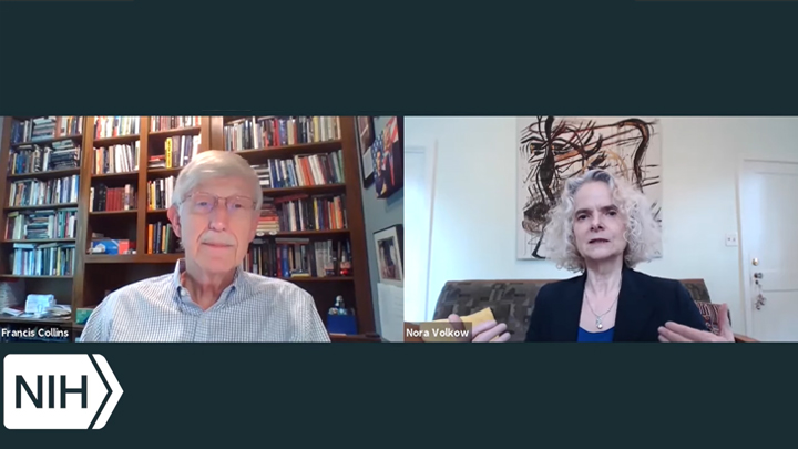
Addressing the Twin Challenges of Substance Use Disorders and COVID-19
Posted on by Dr. Francis Collins
The coronavirus disease 2019 (COVID-19) pandemic is having a wide range of negative impacts on people affected by a variety of health conditions. Among the hardest hit are individuals struggling with substance use disorders, with recent data indicating that suspected drug-related overdoses and deaths are on the rise across the United States [1].
One recent analysis of nationwide surveillance data, collected by the federal Overdose Detection Mapping and Application Program, found that suspected drug overdoses rose by 18 percent in March, 29 percent in April, and 42 percent in May compared to the same months in 2019 [2]. Another analysis of state and local mortality data showed that drug-related deaths have increased about 13 percent so far this year, compared to last year [3].
To find out what may be contributing to this tragic situation and learn what NIH-funded research is doing to help, I recently had a conversation with Dr. Nora Volkow, Director of NIH’s National Institute on Drug Abuse (NIDA). Here’s a condensed version of our interview, which took place via videoconference, with both of us linking in from our homes near NIH’s main campus in Bethesda, MD
Collins: Here we are today talking about two public health crises: the crisis of COVID-19 and another crisis that has been going on for quite some time, of drug overdoses and drug deaths. The opioid crisis is difficult in any circumstance, but when you add to it what’s happening right now with the global COVID-19 pandemic, it becomes difficult squared. What has happened during this pandemic?
Volkow: One of the first things that we’ve heard from the communities and the families afflicted by addiction is that the support systems that were there to help people achieve recovery are no longer present. At the same time, it’s been much harder to get access to some of the treatment programs, including hospital emergency departments that can initiate treatment. It’s also been more difficult to access syringe exchange programs and programs, like Narcotics Anonymous, that provide people with a mentor and a social support system that’s fundamental for recovery. Part of recovery is also for individuals to work at re-building their lives, and that too has become much more challenging due to the threat of COVID-19.
All of these aspects are translating into much more stress. And stress, as we know, is one of the factors that leads people to relapse. Stress is also a factor that leads many to increase the consumption of drugs.
Collins: What about the impact of the stay-at-home orders for people who are depending on social networks? You’ve talked about Narcotics Anonymous as an example. But for anybody who’s faced stress challenges, mental health issues, which often coexist with drug problems, what’s the effect of losing those face-to-face social connections?
Volkow: Isolation is difficult for anyone. We depend on others for our wellbeing. The harder our situation, the more vulnerable we are if we don’t have those support systems.
One of the major concerns that we’ve had all along is not just the enormous risk of relapse in many people, but also the risk of suicide—which is always much higher in individuals that are addicted to drugs, particularly to opioids. Indeed, there’s been an increase in the number of suicides associated with the COVID-19 pandemic, including among people that are addicted.
One of the elements we are using to try to overcome that is virtual interactions, like we are having right now. They are fulfilling, certainly for me. And when we’ve surveyed patients and families to see how much these virtual support systems are helping them, we see in many instances that this can be lifesaving. For example, with telehealth, a physician now can prescribe buprenorphine [a treatment medication] without necessarily having to see the individual physically. That’s a major breakthrough because it expands the number of people that can be treated. So, you can provide buprenorphine, and you can also provide support that someone with co-morbid mental illness may need. It’s not the same as physically being with others, but we have to recognize virtual technologies may enable greater equity in providing treatments.
Collins: What’s happened to methadone clinics, a place where people were required to show up in person every day? What’s become of people who depended on those?
Volkow: These spaces are small and there’s not enough staff, so it was very, very high risk. So, one of the positives of COVID-19 is that there was a change in the policy that enabled a methadone clinic to provide take-home methadone for patients, rather than have them come in daily and often at very restricted times, which made it incredibly difficult to comply.
We’re now trying to evaluate the outcomes when people are given take-home methadone. If we can show from evidence that the outcomes are as good as when you go in daily, then we hope that will help to transform these policies permanently.
Collins: So, there’s a silver lining in a few places. Are people who suffer from drug use disorders at increased risk of getting sick from COVID-19?
Volkow: There are many factors that place them at very, very high risk: pharmacological, structural, and social.
Pharmacological, because these drugs negatively affect multiple systems in your body and one of the main targets is the pulmonary system. If your pulmonary system already has pathology because of prior conditions, it’s much easier for the virus to actually infect you and lead to negative outcomes. That pertains to cigarette smoking that produces COPD and pulmonary damage, as well as to very toxic drugs like methamphetamine, which produces pulmonary hypertension; or opioids, which actually depress respiration and produce hypoxia.
You can see that the combination of depressed respiration and having a viral infection that attacks your lungs is not going to be positive. Indeed, it is very likely that that combination lowers the threshold for people to die from overdoses or to die from COVID-19. Drugs can also affect the cardiovascular system and the metabolic system, so all of the factors that we’ve identified as conditions that make you more vulnerable to COVID-19 are affected by drugs.
Then there are structural issues. We’ve already discussed methadone clinics, which put people together in very close spaces. Before COVID-19, one of our main priorities was to bring the treatment of substance use disorder and the screening into the healthcare system. But now the healthcare system is saturated and individuals who’ve gotten their treatment in healthcare systems no longer can access them and that restricts their ability to seek help. In our country, we basically criminalize people who take drugs, and many of them are in jail systems and prisons, where COVID-19 infections can rapidly occur. That is another element where they are at much higher risk.
Also, the number of individuals with substance use disorder who have medical insurance is much less than that of the general population. Not having such insurance is associated with a greater likelihood of having chronic medical conditions, which again is another risk factor for COVID-19. This mixes the structural with the social and, in the social category, you also have stigma.
Stigmatizing individuals with addiction makes them very vulnerable. That’s because, first of all, they are afraid to seek help—they don’t want to be discriminated against. Secondly, if they are in a situation where decisions are being made about providing medical care when resources are limited, that stigma can make them much more vulnerable.
While we are dealing with COVID-19, we cannot ignore the disparities that exist in our society. This pandemic has made it very clear how horrifically disparate health outcomes are between groups of people in our country.
Collins: Nora, you’ve been a real leader on what we might do to try to bring attention to helping people with drug use problems in the criminal justice system. This is often a point where an opportunity for treatment arises, but unfortunately that opportunity is often missed.
Volkow: One of our priorities as we address the opioid crisis is to do research in justice settings in order to be able to identify the models that lead to the best outcomes and to understand how to implement them. This has resulted in the creation of a research network that enables us to connect across the justice and the healthcare systems.
The network that started to emerge before COVID-19 hit has given us an opportunity to get direct information about what’s happening out there. From what we know, because prisons and jails are at such high risk for infection, many states—if not all—are releasing people that are not violent into their communities. Many of them have a substance use disorder. If someone has a long history of a substance use disorder, you cannot release them into the community without a support system, especially in the midst of the COVID-19 pandemic, where it’s hard to find a job and their families may be rejecting them. You can predict the outcome is going to be very poor, including dying from overdoses.
So, we now have a chance to show that treating these people in their community with appropriate support is going to lead to much better outcomes than leaving them in jail or prison. We are now working with our researchers and with appropriate agencies to figure out how to provide the support that’s necessary as individuals with substance use disorders are released into their communities. It can go both ways. Without support, the outcomes may be very poor. With support, we have the opportunity of transforming the way that we deal with addiction in this country.
Collins: A lot of people may not realize that effective medical treatment for substance use disorders does exist. Treatment has been demonstrated to change lives and improve outcomes over the long term. Still, a lot of folks out there think it’s just hopeless, or, alternatively, if someone just had a little bit more willpower, he or she would be able to take care of this. Please say a little bit about what the current treatment options are, and what the evidence is that they’re needed if you’re going to help somebody recover from a substance use disorder.
Volkow: There are medications for alcoholism and medications for nicotine use disorders. But, by far, the most effective medications are for opioid addiction. It’s very frustrating these medications are not necessarily given to patients—or sometimes even given to patients, but they reject them. I think part of the issue is because of the stigma against the medications. The opioid crisis has helped smooth that out somewhat, so there’s been a greater acceptance of medication. In partnership with the pharmaceutical industry, we have also been working towards developing extended-release formulations that make it much easier for people to take these medications.
In parallel, not just for opioid addiction, we have built up the scientific evidence for behavioral interventions that can improve outcomes for people with substance use disorder in general, if provided concurrently with medical treatment. Recognizing that there is a high risk of comorbidity with mental illness, we also need to provide treatments to address psychiatric disease problems or symptoms, as well as the addiction process. A lot of the work right now is going into creating models that allow this comprehensive treatment, tailored to the needs of the person.
Collins: Where can people who have a family member or friend who’s struggling with substance use disorder in the midst of COVID-19 go to get reliable evidence-based information about treatment programs?
Volkow: They can go to the NIDA website or the website of NIH’s sister agency, the Substance Abuse and Mental Health Services Administration (SAMHSA). One of the problems is that there hasn’t been any way of assessing the quality of treatment for substance use disorder. For many other conditions, you can check the track records of this or that hospital for this or that surgery, but such information does not exist for substance use disorder.
So, we’ve been funding researchers to develop metrics that can predict good outcomes in treatment programs. These metrics can be based on the experiences of people and family that actually took these services, and from the structural characteristics of the program, such as whether they have the evidence-based components shown by research to lead to better outcomes. Researchers are now developing “report cards” for treatment programs that hopefully will do two things: give a family member a sense of how others are rating a program, and, importantly, incentivize treatment programs to do better.
Collins: It would be wonderful to have more objective data for people searching for good answers. Now, let’s talk about HEAL, which stands for Helping to End Addiction Long-term. HEAL is a trans-agency initiative funded by the Congress to support research to address, from multiple different directions, multiple different problems relating to addiction and chronic pain.
How does the HEAL initiative need to adapt to the current health crisis of COVID-19? And what’s your institute doing to try to address some of the significant problems that have emerged in just the last two or three months?
Volkow: COVID-19 has placed HEAL and much of our other research on a very slow trajectory. For example, one program that we were very interested in expanding was the use of the emergency department for the screening of opioid use disorder and the initiation of treatment medications. Another major HEAL program was going to start using the justice system to conduct clinical trials to evaluate the outcomes of different types of medication for opioid use disorder. They are all basically on hold.
Collins: Nora, what’s your hope going forward over the next few months? What can NIH do to try to address this situation in the most effective way possible?
Volkow: I am optimistic because I can see how science can help to solve extremely challenging problems. I think this is the time for science to shine again and show us that methodologies aimed at gathering objective data to develop optimal solutions can resolve problems. But the question is: how long will it take?
I’ve been very impressed about how these devastating circumstances have led us to question the pace at which we moved projects in the past. I think it is wonderful that we have recognized that time is a luxury, that we need to move rapidly. With respect to the issue of substance use disorders, I would hope that, as we as a nation become aware of the suffering that the COVID-19 pandemic is putting on all of us, we become more empathetic to the suffering of others.
And as I see the movements across the country speaking out against injustice, I would hope that this will also extend to diseases that have been stigmatized. We need to modify our stigma so we provide the same level of importance to treating these diseases and supporting people afflicted by them.
I think that science will prevail. What is going to be important is that we also allow for our humanity in order to use that science in a way that everyone can take advantage of it.
Collins: That’s a wonderful way to wind up because I think the calling to bring together science and compassion is what drives all of us who have the privilege of working at NIH, the largest supporter of biomedical research in the world. Our purpose is clear: to find answers for all of these difficult problems that cause suffering and early death for people who deserve better.
Our vision is set on helping the most vulnerable populations right now. COVID-19 has pointed us toward that, and our discussion about those who suffer from substance use disorders also focuses on that.
I’m always one who likes to talk about hope, because, after all, that’s what we get up in the morning thinking about at NIH. We hope that our research efforts are going to lead to a new vaccine or a new treatment for COVID-19, or a better way of helping people who have been afflicted with drug problems.
Yet one of my favorite sayings is that “hope is a privilege that attaches to action.” This means that you can’t just say “Well, I hope for something,” unless you attach that hope to concrete actions you’re going to take.
Nora, your institute has been living that out. You don’t just hope that something good will happen to turn the tide of this terrible crisis of suffering and death from opioid overdoses, you’re all about action. So, thank you for your incredible dedication to the science and to the people whom we are trying to serve.
Volkow: Francis, thanks very much for your support.
References:
{1] Issue brief: Reports of increases in opioid-related overdose and other concerns during COVID pandemic, American Medical Association. Updated July 20, 2020
[2] “Cries for help’: Drug overdoses are soaring during the coronavirus pandemic.” William Wan, Healther Long. The Washington Post, July 1, 2020.
[3] “In the shadow of the pandemic, U.S. drug overdose deaths resurge to record.” Josh Katz, Abby Goodnough, Margot Sanger-Katz. July 15, 2020.
Links:
Coronavirus (COVID-19) (NIH)
Overdose Mapping Application Program (White House Office of National Drug Control Policy, Washington, D.C.)
Helping to End Addiction Long-term (HEAL) Initiative (NIH)
National Institute on Drug Abuse (NIH)
Video: Effects of COVID-19 on the Opioid Crisis: Francis Collins with Nora Volkow (National Institute on Drug Abuse/NIH)
Substance Abuse and Mental Health Services Administration (SAMHSA)
NIH’s All of Us Program Joins Fight Against COVID-19
Posted on by Dr. Francis Collins

We’ve learned so much about coronavirus disease 2019 (COVID-19), but there’s still much more that we need to learn in order to defeat this devastating pandemic. Among the critical questions: why do some young people who appear healthy and have no history of chronic disease get very sick from the virus? And why do some people in their 80s or 90s seemingly just shrug off the infection? There’s something going on biologically, but we don’t yet have the answers.
We do, however, have some resources that will enable us to examine lots of data in search of biological clues. One of them is NIH’s All of Us Research Program, which is seeking the help of 1 million people to build one of the most diverse health databases in our nation’s history. Two years after its national launch, the program already has enrolled nearly 350,000 diverse participants from across the United States.
As its name suggests, All of Us is open to all people over age 18 in communities all around the country. An important strength of the effort has been welcoming participants from all backgrounds. Indeed, about 75 percent of people who have volunteered for the program come from groups that have traditionally been underrepresented in medical research. That includes people from many racial and ethnic minority groups, as well as those of many different ages, socioeconomic backgrounds, and geographic locations, including remote and rural areas.
Because of COVID-19 and the need for physical distancing to curb the spread of the potentially deadly virus, All of Us has been forced to halt temporarily all in-person appointments. But program leaders, including Josh Denny, chief executive officer of All of Us, and Kelly Gebo, the program’s chief medical and scientific officer, saw an opportunity to roll up their sleeves and help during this unprecedented public health challenge. In fact, Gebo reports that they’d already been hearing from many of their participant partners that they wanted to be a part of the solution to the COVID-19 pandemic.
To rise to this challenge, the All of Us Research Program has just announced three initiatives to assist the scientific community in seeking new insights into COVID-19. The program will:
• Test blood samples from 10,000 or more participants for the presence of SARS-CoV-2 antibodies, indicating prior infection. The testing will start on samples collected in March 2020 and work backward until positive tests are no longer found. This will show the prevalence of novel coronavirus exposure among All of Us participants from across the country, allowing researchers to sift through the data and assess the varying rates and timing of infections across regions and communities.
• Rapidly collect relevant information from more than 200,000 participants who have shared their electronic health records. A number of those participants have already either been diagnosed with COVID-19 or sought health care for related symptoms. The program is working to standardize this information. It will help researchers look for patterns and learn more about COVID-19 symptoms and associated health problems, as well as the effects of different medicines and treatments.
• Deploy a new online survey to understand better the effects of the COVID-19 pandemic on participants’ physical and mental health. This 20- to 30-minute survey is designed both for participants who have been ill with COVID-19 and those who have not knowingly been infected. Questions will be included on COVID-19 symptoms, stress, social distancing and the economic impacts of the pandemic. Participants are invited to take the survey each month until the pandemic ends, so researchers can study the effects of COVID-19 over time and begin to better understand how and why COVID-19 affects people differently.
As this data becomes available, researchers will look for new leads to inform our efforts to bring greater precision to the diagnosis, treatment, and prevention of COVID-19, including for those communities that have been hit the hardest. Another hope is that what is learned about COVID-19 through All of Us and other NIH-supported research will provide us with the knowledge and tools we need to avert future pandemics,
In case you’re wondering, I happen to be among the thousands of people who’ve already volunteered to take part in All of Us. If you’d like to get involved too, new participants are always welcome to join.
Links:
Coronavirus (COVID-19) (NIH)
All of Us Research Program (NIH)
Join All of Us (NIH)
Next Page