2016 May
Gene Duplication: New Analysis Shows How Extra Copies Split the Work
Posted on by Dr. Francis Collins
 The human genome contains more than 20,000 protein-coding genes, which carry the instructions for proteins essential to the structure and function of our cells, tissues and organs. Some of these genes are very similar to each other because, as the genomes of humans and other mammals evolve, glitches in DNA replication sometimes result in extra copies of a gene being made. Those duplicates can be passed along to subsequent generations and, on very rare occasions, usually at a much later point in time, acquire additional modifications that may enable them to serve new biological functions. By starting with a protein shape that has already been fine-tuned for one function, evolution can produce a new function more rapidly than starting from scratch.
Pretty cool! But it leads to a question that’s long perplexed evolutionary biologists: Why don’t duplicate genes vanish from the gene pool almost as soon as they appear? After all, instantly doubling the amount of protein produced in an organism is usually a recipe for disaster—just think what might happen to a human baby born with twice as much insulin or clotting factor as normal. At the very least, duplicate genes should be unnecessary and therefore vulnerable to being degraded into functionless pseudogenes as new mutations arise over time
An NIH-supported team offers a possible answer to this question in a study published in the journal Science. Based on their analysis of duplicate gene pairs in the human and mouse genomes, the researchers suggest that extra genes persist in the genome because of rapid changes in gene activity. Instead of the original gene producing 100 percent of a protein in the body, the gene duo quickly divvies up the job [1]. For instance, the original gene might produce roughly 50 percent and its duplicate the other 50 percent. Most importantly, organisms find the right balance and the duplicate genes can easily survive to be passed along to their offspring, providing fodder for continued evolution.
Creative Minds: Breaking Size Barriers in Cryo-Electron Microscopy
Posted on by Dr. Francis Collins

Dmitry Lyumkis
When Dmitry Lyumkis headed off to graduate school at The Scripps Research Institute, La Jolla, CA, he had thoughts of becoming a synthetic chemist. But he soon found his calling in a nearby lab that imaged proteins using a technique known as single-particle cryo-electron microscopy (EM). Lyumkis was amazed that the team could take a purified protein, flash-freeze it in liquid nitrogen, and then fire electrons at the protein, capturing the resulting image with a special camera. Also amazing was the sophisticated computer software that analyzed the raw 2D camera images, merging the data and reconstructing it into 3D representations of the protein.
The work was profoundly complex, but Lyumkis thrives on solving extremely difficult puzzles. He joined the Scripps lab to become a structural biologist and a few years later used single-particle cryo-EM to help determine the atomic structure of a key protein on the surface of the human immunodeficiency virus (HIV), the cause of AIDS. The protein had been considered one of the greatest challenges in structural biology and a critical target in developing an AIDS vaccine [1].
Now, Lyumkis has plans to take single-particle cryo-EM to a whole new level—literally. He wants to develop new methods that allow it to model the atomic structures of much smaller proteins. Right now, single-particle cryo-EM has worked with proteins as small as roughly 150 kilodaltons, a measure of a protein’s molecular weight (the approximate average mass of a protein is 53 kDa). Lyumkis plans to drop that number well below 100 kDa, noting that if his new methods work as he hopes, there should be very little, if any, lower size limit to get the technique to work. He envisions generating within a matter of days or weeks the precise structure of an average-sized protein involved in a disease, and then potentially handing it off as an atomic model for drug developers to target for more effective treatment.
Alzheimer’s Disease: Tau Protein Predicts Early Memory Loss
Posted on by Dr. Francis Collins
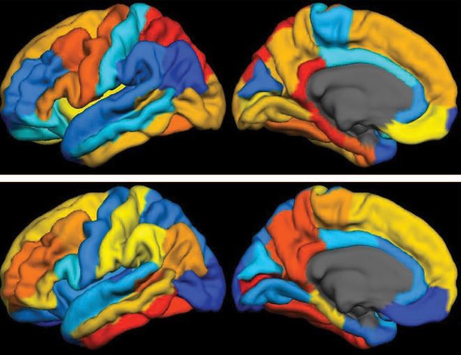
Caption: PET scan images show distribution of tau (top panel) and beta-amyloid (bottom panel) across a brain with early Alzheimer’s disease. Red indicates highest levels of protein binding, dark blue the lowest, yellows and oranges indicate moderate binding.
Credit: Brier et al., Sci Transl Med
In people with Alzheimer’s disease, changes in the brain begin many years before the first sign of memory problems. Those changes include the gradual accumulation of beta-amyloid peptides and tau proteins, which form plaques and tangles that are considered hallmarks of the disease. While amyloid plaques have received much attention as an early indicator of disease, until very recently there hadn’t been any way during life to measure the buildup of tau protein in the brain. As a result, much less is known about the timing and distribution of tau tangles and its relationship to memory loss.
Now, in a study published in Science Translational Medicine, an NIH-supported research team has produced some of the first maps showing where tau proteins build up in the brains of people with early Alzheimer’s disease [1]. The new findings suggest that while beta-amyloid remains a reliable early sign of Alzheimer’s disease, tau may be a more informative predictor of a person’s cognitive decline and potential response to treatment.
Snapshots of Life: Portrait of Zika Virus
Posted on by Dr. Francis Collins
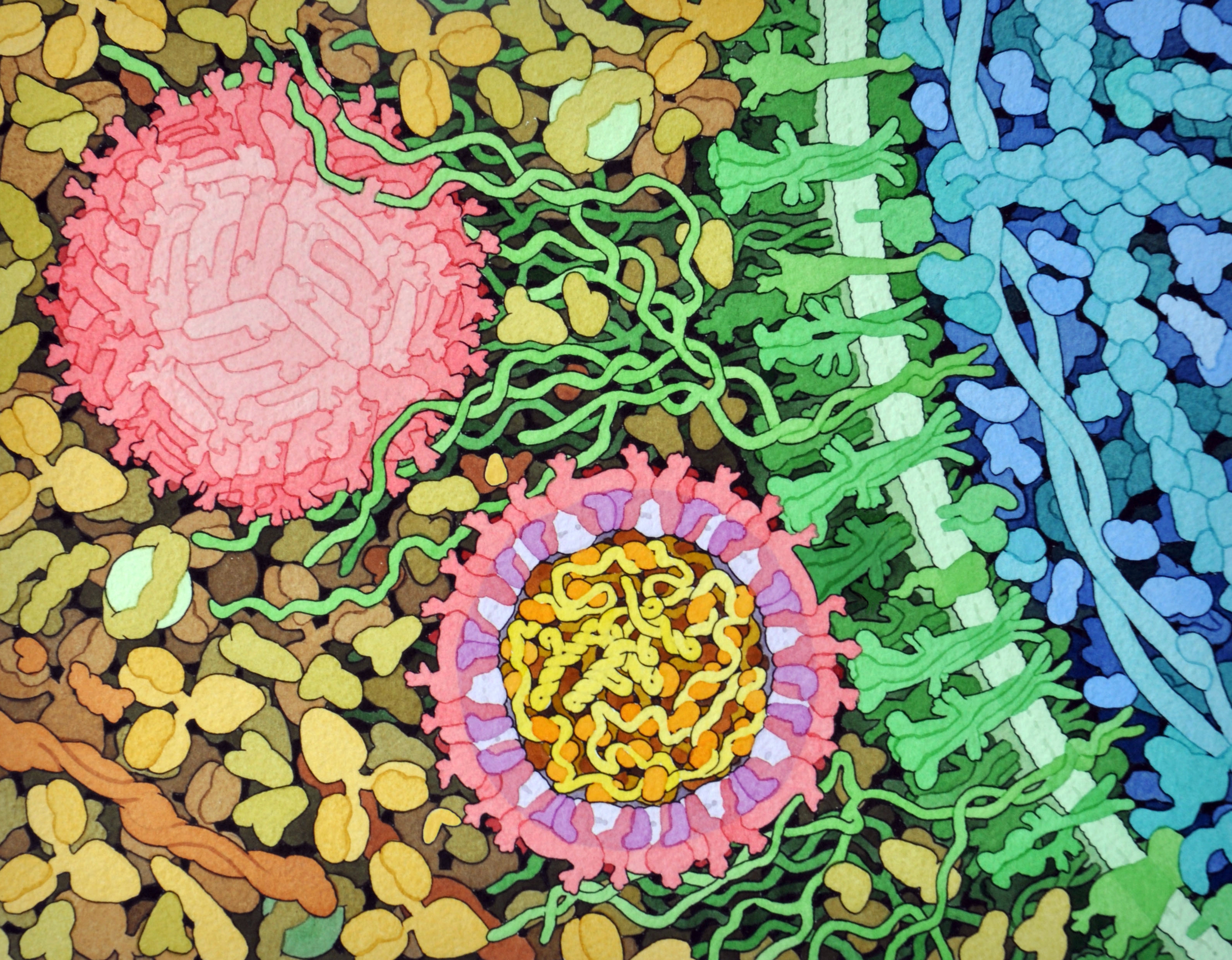
Credit: David Goodsell, The Scripps Research Institute
This lively interplay of shape and color is an artistic rendering of the Zika virus preparing to enter a cell (blue) by binding to its protein receptors (green). The spherical structures (pink) represent two Zika viruses in a blood vessel filled with blood plasma cells (tan). The virus in the middle in cross section shows viral envelope proteins (red) studding the outer surface, with membrane proteins (pink) embedded in a fatty layer of lipids (light purples). In the innermost circle, you can see the viral genome (yellow) coiled around capsid proteins (orange).
This image was sketched and hand-painted with watercolors by David Goodsell, a researcher and illustrator at The Scripps Research Institute, La Jolla, CA. Goodsell put paint and science to paper as part of the “Molecule of the Month” series run by RCSB Protein Data Bank (PDB), which NIH co-supports with the National Science Foundation and the Department of Energy. The PDB, which contains structural data on thousands of proteins and small molecules, uses its “Molecule of the Month” series to help students visualize a molecule or virus and to encourage their exploration of structural biology and its applications to medicine.
Portable System Uses Light to Diagnose Bacterial Infections Faster
Posted on by Dr. Francis Collins

Caption: PAD system. Left, four optical testing cubes (blue and white) stacked on the electronic base station (white with initials); right, a smartphone with a special app to receive test results transmitted by the electronic base station.
Credit: Park et al. Sci. Adv. 2016
Every year, hundreds of thousands of Americans acquire potentially life-threatening bacterial infections while in the hospital, nursing home, or other health-care settings [1]. Such infections can be caused by a variety of bacteria, which may respond quite differently to different antibiotics. To match a patient with the most appropriate antibiotic therapy, it’s crucial to determine as quickly as possible what type of bacteria is causing his or her infection. In an effort to improve that process, an NIH-funded team is working to develop a point-of-care system and smartphone app aimed at diagnosing bacterial infections in a faster, more cost-effective manner.
The portable new system, described recently in the journal Science Advances, uses a novel light-based method for detecting telltale genetic sequences from bacteria in bodily fluids, such as blood, urine, or drainage from a skin abscess. Testing takes place within small, optical cubes that, when placed on an electronic base station, deliver test results within a couple of hours via a simple readout sent directly to a smartphone [2]. When the system was tested on clinical samples from a small number of hospitalized patients, researchers found that not only did it diagnose bacterial infections about as accurately and more swiftly than current methods, but it was also cheaper. This new system can potentially also be used to test for the presence of antibiotic-resistant bacteria and contamination of medical devices.
Creative Minds: Considering the Social Determinants of Health
Posted on by Dr. Francis Collins

Sanjay Basu
When Sanjay Basu was growing up in Arizona in the 1980s, his mother contracted a devastating lung infection known as valley fever. Caused by a fungus (called Coccidioides) common in the southwest United States, the condition often affects construction or agricultural workers who inhale the fungal spores while working the soil. Basu’s mother didn’t work in agriculture or construction, but the family did happen to live near a construction site. She spent about nine years in and out of intensive care units battling her illness. She survived, but still has difficulty breathing.
This wrenching experience gave Basu a first-hand appreciation for the social determinants of health—the conditions in which people live and the myriad internal and external forces that dynamically shape them. Now an assistant professor at Stanford University, Palo Alto, CA, Basu has dedicated his career to studying the social determinants of health disparities, health differences that adversely affect disadvantaged populations. He recently received an NIH Director’s New Innovator Award to examine U.S. social assistance programs and their effects on a range of health outcomes over the last 40-plus years. He’ll consider eight federal and state programs—including income, housing, and food assistance programs—that reach more than 1 in 3 Americans.
Fighting Depression: Ketamine Metabolite May Offer Benefits Without the Risks
Posted on by Dr. Francis Collins

Thinkstock/Ryan McVay
For people struggling with severe depression, antidepressants have the potential to provide much-needed relief, but they often take weeks to work. That’s why there is growing excitement about reports that the anesthetic drug ketamine, when delivered intravenously in very low doses, can lift depression and suicidal thoughts within a matter of hours. Still, there has been reluctance to consider ketamine for widespread treatment of depression because, even at low doses, it can produce very distressing side effects, such as dissociation—a sense of disconnection from one’s own thoughts, feelings, and sense of identity. Now, new findings suggest there may be a way to tap into ketamine’s depression-fighting benefits without the side effects.
In a mouse study published in the journal Nature, an NIH-funded research team found that the antidepressant effects of ketamine are produced not by the drug itself, but by one of its metabolites—a substance formed as the body breaks ketamine down. What’s more, the work demonstrates that this beneficial metabolite does not cause the risky dissociation effects associated with ketamine. While further development and subsequent clinical trials are needed, the findings are a promising step toward the development of a new generation of rapid-acting antidepressant drugs.
Cool Videos: Another Kind of Art Colony
Posted on by Dr. Francis Collins
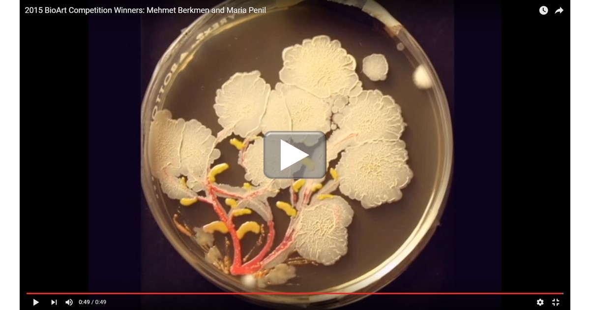 As long as researchers have been growing bacteria on Petri dishes using a jelly-like growth medium called agar, they have been struck by the interesting colors and growth patterns that microbes can produce from one experiment to the next. In the 1920s, Sir Alexander Fleming, the Scottish biologist who discovered penicillin, was so taken by this phenomenon that he developed his own palette of bacterial “paints” that he used in his spare time to create colorful pictures of houses, ballerinas, and other figures on the agar [1].
Fleming’s enthusiasm for agar art lives on among the current generation of microbiologists. In this short video, the agar (yellow) is seeded with bacterial colonies and, through the magic of time-lapse photography, you can see the growth of the colonies into what appears to be a lovely bouquet of delicate flowers. This piece of living art, developing naturally by bacterial colony expansion over the course of a week or two, features members of three bacterial genera: Serratia (red), Bacillus (white), and Nesterenkonia (light yellow).
A Look Inside a Beating Heart Cell
Posted on by Dr. Francis Collins
Caption: Microtubules (blue) in a beating heart muscle cell, or cardiomyocyte. Credit: Lab of Ben Prosser, Ph.D., Perelman School of Medicine, University of PennsylvaniaYou might expect that scientists already know everything there is to know about how a healthy heart beats. But researchers have only recently had the tools to observe some of the dynamic inner workings of heart cells as they beat. Now an NIH-funded team has captured video to show that a component of a heart muscle cell called microtubules—long thought to be very rigid—serve an unexpected role as molecular shock absorbers.
As described for the first time recently in the journal Science, the microtubules buckle under the force of each contraction of the muscle cell before springing back to their original length and form. The team also details a biochemical process that allows a cell to fine-tune the level of resistance that the microtubules provide. The findings have important implications for understanding not only the mechanics of a healthy beating heart, but how the abnormal stiffening of heart cells might play a role in various forms of cardiac disease.