heart muscle cells
A Look Inside a Beating Heart Cell
Posted on by Dr. Francis Collins
Caption: Microtubules (blue) in a beating heart muscle cell, or cardiomyocyte. Credit: Lab of Ben Prosser, Ph.D., Perelman School of Medicine, University of PennsylvaniaYou might expect that scientists already know everything there is to know about how a healthy heart beats. But researchers have only recently had the tools to observe some of the dynamic inner workings of heart cells as they beat. Now an NIH-funded team has captured video to show that a component of a heart muscle cell called microtubules—long thought to be very rigid—serve an unexpected role as molecular shock absorbers.
As described for the first time recently in the journal Science, the microtubules buckle under the force of each contraction of the muscle cell before springing back to their original length and form. The team also details a biochemical process that allows a cell to fine-tune the level of resistance that the microtubules provide. The findings have important implications for understanding not only the mechanics of a healthy beating heart, but how the abnormal stiffening of heart cells might play a role in various forms of cardiac disease.
Snapshots of Life: Mending Broken Hearts
Posted on by Dr. Francis Collins
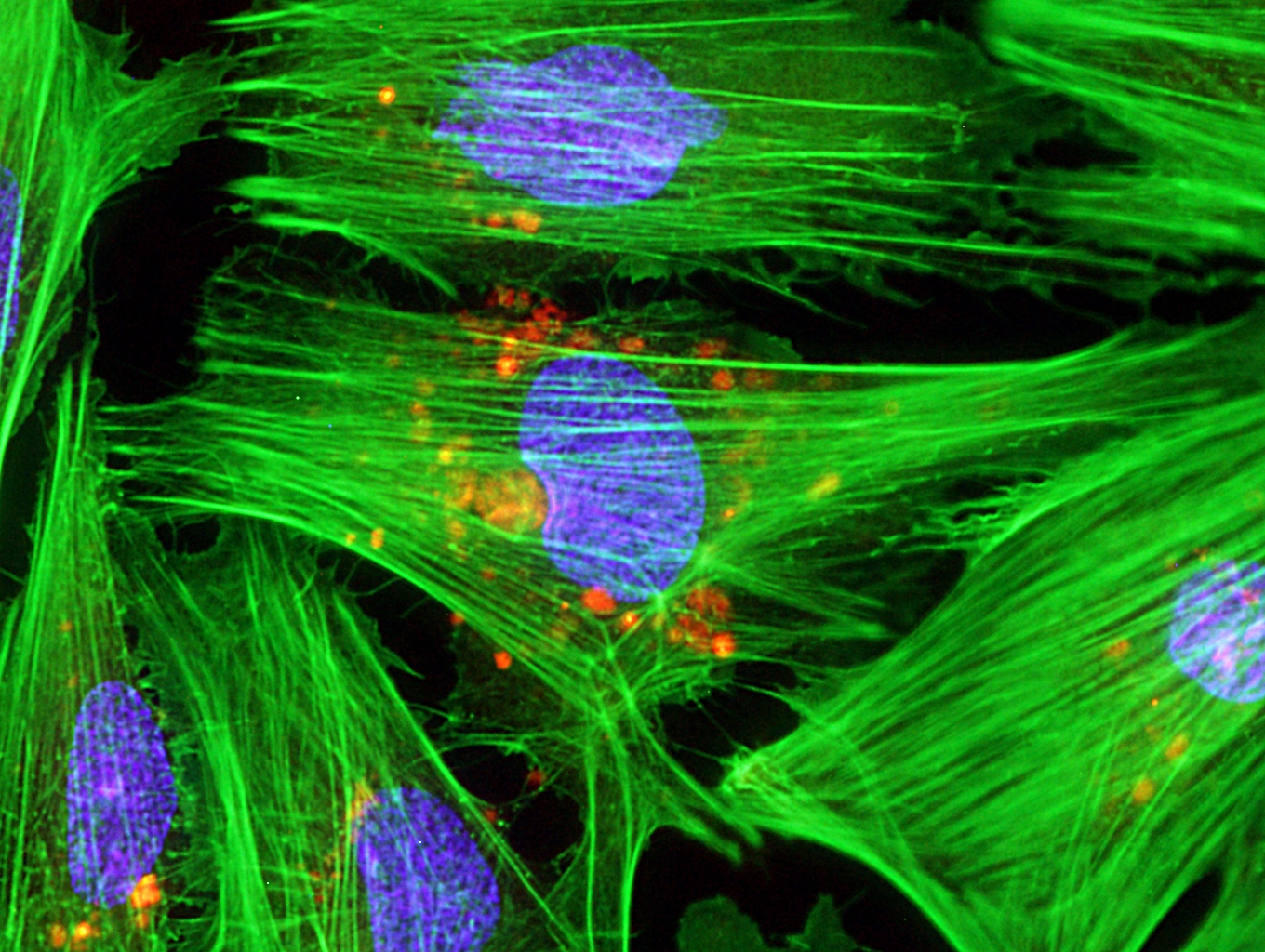
Caption: Micrograph of laboratory-grown rat heart muscle cells. Fluorescent labeling shows mitochondria (red), cytoskeleton (green), and nuclei (blue).
Credit: Credit: Douglas B. Cowan and James D. McCully, Harvard Medical School, Boston
This may not look like your average Valentine’s Day card, but it’s an image sure to warm the hearts of many doctors and patients. Why? This micrograph, a winner in the Federation of American Societies for Experimental Biology’s 2013 BioArt Competition, shows cells that have been specially engineered to repair the damage done by heart attacks—which strike more than 700,000 Americans every year.
Working with rat heart muscle cells grown in a lab dish, NIH-supported bioengineers at Harvard Medical School used transplant techniques to boost the number of tiny powerhouses, called mitochondria, within the cells. If you look closely at the image above, you’ll see the heart muscle cells are tagged in green, their nuclei in blue, and their mitochondria in red.