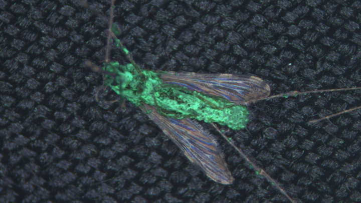
mosquito-borne illnesses
3D Animation Captures Viral Infection in Action
Posted on by Lawrence Tabak, D.D.S., Ph.D.
With the summer holiday season now in full swing, the blog will also swing into its annual August series. For most of the month, I will share with you just a small sampling of the colorful videos and snapshots of life captured in a select few of the hundreds of NIH-supported research labs around the country.
To get us started, let’s turn to the study of viruses. Researchers now can generate vast amounts of data relatively quickly on a virus of interest. But data are often displayed as numbers or two-dimensional digital images on a computer screen. For most virologists, it’s extremely helpful to see a virus and its data streaming in three dimensions. To do so, they turn to a technological tool that we all know so well: animation.
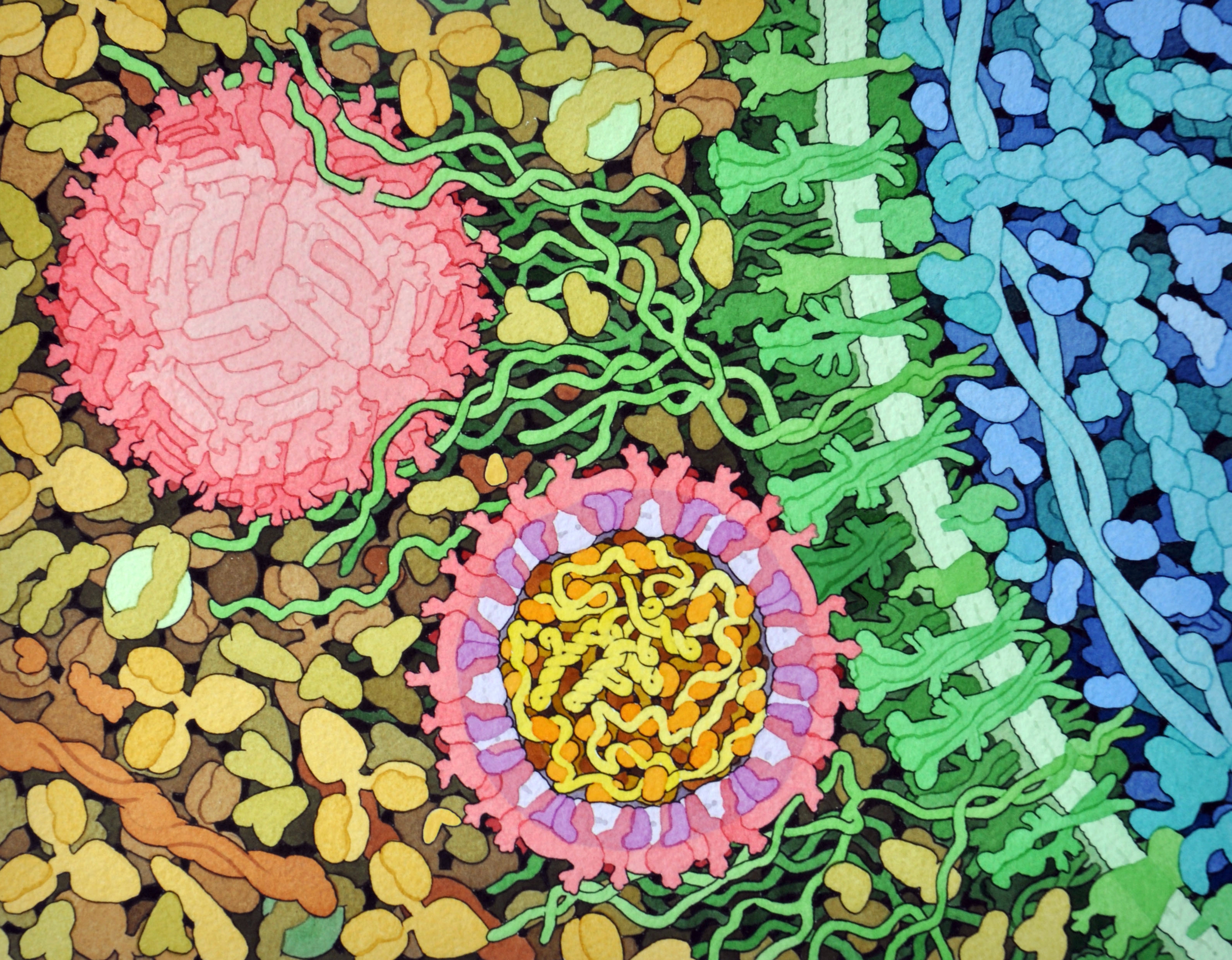
This research animation features the chikungunya virus, a sometimes debilitating, mosquito-borne pathogen transmitted mainly in developing countries in Africa, Asia and the Americas. The animation illustrates large amounts of research data to show how the chikungunya virus infects our cells and uses its specialized machinery to release its genetic material into the cell and seed future infections. Let’s take a look.
In the opening seconds, you see how receptor binding glycoproteins (light blue), which are proteins with a carbohydrate attached on the viral surface, dock with protein receptors (yellow) on a host cell. At five seconds, the virus is drawn inside the cell. The change in the color of the chikungunya particle shows that it’s coated in a vesicle, which helps the virus make its way unhindered through the cytoplasm.
At 10 seconds, the virus then enters an endosome, ubiquitous bubble-like compartments that transport material from outside the cell into the cytosol, the fluid part of the cytoplasm. Once inside the endosome, the acidic environment makes other glycoproteins (red, blue, yellow) on the viral surface change shape and become more flexible and dynamic. These glycoproteins serve as machinery that enables them to reach out and grab onto the surrounding endosome membrane, which ultimately will be fused with the virus’s own membrane.
As more of those fusion glycoproteins grab on, fold back on themselves, and form into hairpin-like shapes, they pull the membranes together. The animation illustrates not only the changes in protein organization, but the resulting effects on the integrity of the membrane structures as this dynamic process proceeds. At 53 seconds, the viral protein shell, or capsid (green), which contains the virus’ genetic instructions, is released back out into the cell where it will ultimately go on to make more virus.
This remarkable animation comes from Margot Riggi and Janet Iwasa, experts in visualizing biology at the University of Utah’s Animation Lab, Salt Lake City. Their data source was researcher Kelly Lee, University of Washington, Seattle, who collaborated closely with Riggi and Iwasa on this project. The final product was considered so outstanding that it took the top prize for short videos in the 2022 BioArt Awards competition, sponsored by the Federation of American Societies for Experimental Biology (FASEB).
The Lee lab uses various research methods to understand the specific shape-shifting changes that chikungunya and other viruses perform as they invade and infect cells. One of the lab’s key visual tools is cryo-electron microscopy (Cryo-EM), specifically cryo-electron tomography (cryo-ET). Cryto-ET enables complex 3D structures, including the intermediate state of biological reactions, to be captured and imaged in remarkably fine detail.
In a study in the journal Nature Communications [1] last year, Lee’s team used cryo-ET to reveal how the chikungunya virus invades and delivers its genetic cargo into human cells to initiate a new infection. While Lee’s cryo-ET data revealed stages of the virus entry process and fine structural details of changes to the virus as it enters a cell and starts an infection, it still represented a series of snapshots with missing steps in between. So, Lee’s lab teamed up with The Animation Lab to help beautifully fill in the gaps.
Visualizing chikungunya and similar viruses in action not only makes for informative animations, it helps researchers discover better potential targets to intervene in this process. This basic research continues to make progress, and so do ongoing efforts to develop a chikungunya vaccine [2] and specific treatments that would help give millions of people relief from the aches, pains, and rashes associated with this still-untreatable infection.
References:
[1] Visualization of conformational changes and membrane remodeling leading to genome delivery by viral class-II fusion machinery. Mangala Prasad V, Blijleven JS, Smit JM, Lee KK. Nat Commun. 2022 Aug 15;13(1):4772. doi: 10.1038/s41467-022-32431-9. PMID: 35970990; PMCID: PMC9378758.
[2] Experimental chikungunya vaccine is safe and well-tolerated in early trial, National Institute of Allergy and Infectious Diseases news release, April 27, 2020.
Links:
Chikungunya Virus (Centers for Disease Control and Prevention, Atlanta)
Global Arbovirus Initiative (World Health Organization, Geneva, Switzerland)
The Animation Lab (University of Utah, Salt Lake City)
Video: Janet Iwasa (TED Speaker)
Lee Lab (University of Washington, Seattle)
BioArt Awards (Federation of American Societies for Experimental Biology, Rockville, MD)
NIH Support: National Institute of General Medical Sciences; National Institute of Allergy and Infectious Diseases
Uncovering a Hidden Zika Outbreak in Cuba
Posted on by Dr. Francis Collins

When Brazilian health officials discovered four years ago that the mosquito-borne Zika virus could cause severe birth defects and other serious health problems, it prompted a major effort across the Americas to curb the infection by controlling mosquitoes and issuing travel advisories. By mid-2017, the hard work seemed to have paid off, and reports of new Zika infections had nearly stopped.
But it turns out Zika may be tougher to control than once thought. New research shows that a large, previously hidden outbreak of Zika virus disease occurred in Cuba, just when it looked like the worst of the epidemic was over. The finding suggests that the Zika virus can linger over long periods, and that mosquito control efforts alone may slow, but not necessarily stop, the march of this potentially devastating infectious disease.
When combating global epidemics, it’s critical to track the spread of dangerous viruses from one place to the next. But some viruses can be tougher to monitor than others, and that certainly has been the case with Zika in the Americas. Though the virus can harm unborn children, many people infected with Zika never feel lousy enough to go to the doctor. Those who do often have symptoms that overlap with other prevalent tropical diseases, such as dengue and chikungunya fever, making it hard to recognize Zika.
That’s why in Brazil, where Zika arrived in the Americas by early 2014, this unexpected viral intruder went undetected for well over a year. By then, it had spread unnoticed to Honduras, circulating rapidly to other Central American nations and Mexico—likely by late 2014 and into 2015.
In the United States, even with close monitoring, a small local outbreak of Zika virus in Florida also went undetected for about three months in 2016 [1]. Then, in 2017, Florida officials began noticing something strange: new cases of Zika infection in people who had traveled to Cuba.
This came as a real surprise because Cuba, unlike most other Caribbean islands, was thought to have avoided an outbreak. What’s more, by then the Zika epidemic in the Americas had slowed to a trickle, prompting the World Health Organization to delist it as a global public health emergency of international concern.
Given the Cuban observation, some wondered whether the Zika epidemic in the Americas was really over. Among them was an NIH-supported research team, including Nathan Grubaugh, Yale School of Public Health, New Haven, CT; Sharon Isern and Scott Michael, Florida Gulf Coast University, Fort Myers; and Kristian Andersen, The Scripps Research Institute, La Jolla, CA, who worked closely with the Florida Department of Health, including Andrea Morrison.
As published in Cell, the team was able to document a previously unreported outbreak in Cuba after the epidemic had seemingly ended [2]. Interestingly, another research group in Spain also recently made a similar observation about Zika in Cuba [3].
In the Cell paper, the researchers show that between June 2017 and October 2018, all but two of 155 cases—a whopping 98 percent of travel-associated Zika infections—traced back to Cuba. Further analysis suggests that the outbreak in Cuba was likely of similar magnitude to outbreaks that occurred in other Caribbean nations.
Their estimates suggest there were likely many thousands of Zika cases in Cuba, and more than 5,000 likely should have been diagnosed and reported in 2017. The only difference was the timing. The Cuban outbreak of Zika virus occurred about a year after infections subsided elsewhere in the Caribbean.
To fill in more of the blanks, the researchers relied on Zika virus genomes from nine infected Florida travelers who returned from Cuba in 2017 and 2018. The sequencing data support multiple introductions of Zika virus to Cuba from other Caribbean islands in the summer of 2016.
The outbreak peaked about a year after the virus made its way to Cuba, similar to what happened in other places. But the Cuban outbreak was likely delayed by a year thanks to an effective mosquito control campaign by local authorities, following detection of the Brazilian outbreak. While information is lacking, including whether Zika infections had caused birth defects, it’s likely those efforts were relaxed once the emergency appeared to be over elsewhere in the Caribbean, and the virus took hold.
The findings serve as yet another reminder that the Zika virus—first identified in the Zika Forest in Uganda in 1947 and for many years considered a mostly inconsequential virus [4]—has by no means been eliminated. Indeed, such unrecognized and delayed outbreaks of Zika raise the risk of travelers innocently spreading the virus to other parts of the world.
The encouraging news is that, with travel surveillance data and genomic tools —enabled by open science—it is now possible to detect such outbreaks. By combining resources and data, it will be possible to develop even more effective and responsive surveillance frameworks to pick up on emerging health threats in the future.
In the meantime, work continues to develop a vaccine for the Zika virus, with more than a dozen clinical trials underway that pursue a variety of vaccination strategies. With the Zika pandemic resolved in the Americas, these studies can be harder to conduct, since proof of efficacy is not possible without active infections. But, as this paper shows, we must remain ready for future outbreaks of this unique and formidable virus.
References:
[1] Genomic epidemiology reveals multiple introductions of Zika virus into the United States. Grubaugh et al. Nature. 2017 Jun 15;546(7658):401-405.
[2] Travel surveillance and genomics uncover a hidden Zika outbreak during the waning epidemic. Grubaugh ND, Saraf S, Gangavarapu K, Watts A, Tan AL, Oidtman RJ, Magnani DM, Watkins DI, Palacios G, Hamer DH; GeoSentinel Surveillance Network, Gardner LM, Perkins TA, Baele G, Khan K, Morrison A, Isern S, Michael SF, Andersen .KG, et. al. Cell. 2019 Aug 22;178(5):1057-1071.e11.
[3] Mirroring the Zika epidemics in Cuba: The view from a European imported diseases clinic. Almuedo-Riera A, Rodriguez-Valero N, Camprubí D, Losada Galván I, Zamora-Martinez C, Pousibet-Puerto J, Subirà C, Martinez MJ, Pinazo MJ, Muñoz J. Travel Med Infect Dis. 2019 Jul – Aug;30:125-127.
[4] Pandemic Zika: A Formidable Challenge to Medicine and Public Health. Morens DM, Fauci AS. J Infect Dis. 2017 Dec 16;216(suppl_10):S857-S859.
Links:
Video: Uncovering Hidden Zika Outbreaks (Florida Gulf Coast University, Fort Myers)
Zika Virus (National Institute of Allergy and Infectious Diseases/NIH)
Zika Virus Vaccines (NIAID)
Zika Free Florida (Florida Department of Health, Tallahassee)
Grubaugh Lab (Yale School of Public Health, New Haven, CT)
Andersen Lab (The Scripps Research Institute, La Jolla, CA)
NIH Support: National Institute of Allergy and Infectious Diseases; National Center for Advancing Translational Sciences
Combating Mosquitoes with an Engineered Fungus
Posted on by Dr. Francis Collins
Almost everywhere humans live on this planet, mosquitoes carry microbes that cause potentially deadly diseases, from West Nile virus to malaria. While chemical insecticides offer a line of defense, mosquito populations often grow resistant to them. So, it’s intriguing to learn that we may now have another ally in this important fight: a genetically engineered fungus!
Reporting in the journal Science, an international research team supported by NIH describes how this new approach might be used to combat malaria [1]. A fungus called Metarhizium pingshaense is a natural enemy of the mosquito, but, by itself, it kills mosquitoes too slowly to control transmission of malaria. To make this fungus an even more efficient mosquito killer, researchers engineered it to carry a gene encoding a toxin, derived from a spider, that is deadly to insects. Tests of the souped-up fungus in a unique contained facility designed to simulate a West African village found it safely and rapidly killed insecticide-resistant mosquitoes, reducing their numbers by more than 99 percent within 45 days.
Mosquitoes are the deadliest animals in the world. More than 3.2 billion people—about half of all humans—are at risk for malaria, and more than 400,000 die each year from the disease. Other mosquito-borne illnesses, including Zika and dengue viruses, sicken millions more each year. By combining existing insect control strategies with the latest technical innovation, it should be possible to lower those numbers.
In the latest study, Raymond St. Leger and Brian Lovett, University of Maryland, College Park, teamed with Abdoulaye Diabate and colleagues from Institut de Recherche en Sciences de la Santé/Cente Muraz, Burkina Faso, West Africa. The researchers employed a strategy that’s been in use around the world for more than 100 years to control agricultural pests.
The approach involves the fungal species Metarhizium, which kills a variety of insects. Earlier studies had shown that spores from a specific Metarhizium strain could make a big enough dent in a mosquito population to raise the possibility of using the fungus to reduce infective bites among humans [2]. But killing off the mosquitoes required very large quantities of fungal spores and usually took a couple of weeks.
Here’s where things turned innovative. To boost the fungus’s potency, St. Leger and colleagues used genetic engineering to add a toxin derived from the Australian Blue Mountains funnel-web spider. The toxin came with a major advantage: the U.S. Environmental Protection Agency (EPA) already has approved its use as a safe-and-effective insecticidal protein.
Besides giving the engineered fungus that ability to produce a spider toxin, the researchers added another clever element. They didn’t want the fungus to produce the toxin all the time—only after it comes in contact with a mosquito’s hemolymph, the insect equivalent of blood. So, they needed to insert a control switch, and the researchers knew just where to find the needed part.
Once inside a mosquito, the fungus naturally produces a structural protein called collagen that shields it from the insect’s immune system. A genetic switch that turns “on” when it detects an insect’s hemolymph controls that collagen production. To ensure that the spider toxin was produced at just the right time, the researchers hotwired their Metarhizium to begin producing it under the control of this same genetic switch.
The next step was to test this modified organism in a more natural, but controlled, environment. The researchers spent more than a year in Burkina Faso building a specialized facility called a MosquitoSphere. It’s similar to a very large greenhouse, but with mosquito netting instead of glass.
The MosquitoSphere has six separate compartments, four of which contain West African huts, along with native plants and breeding sites for mosquitoes. The researchers hung a black cotton sheet, previously soaked in sesame oil, on the wall of a hut in each of three chambers.
In one hut, the sesame oil contained genetically engineered fungal spores. In the second hut, the oil contained natural fungal spores. In the third hut, there were no spores at all. Then, they released 1,000 adult male and 500 adult female mosquitoes into each chamber and watched what happened over the next 45 days.
In the hut without spores, the mosquitoes established a stable population of almost 1,400. In the chamber with the natural spores, 450 mosquitoes survived. But, in the chamber with the engineered fungus, the researchers counted just 13 survivors—too few to sustain a viable population.
The researchers say they suspect the fungus would be relatively easy to contain in nature. It’s sticky and not easily airborne. The spores are also extremely sensitive to sunlight, making it difficult for them to travel far. Importantly, the fungus didn’t harm other beneficial insects, including honeybees.
Caution is warranted before considering the release of a genetically engineered organism into the wild. In the meantime, the genetically engineered fungus also will serve as a platform for continued technology development.
The system can be readily adapted to target mosquitoes or other insects , perhaps using different natural toxins if insects might grow resistant to Metarhizium just as they have to traditional insecticides. Interestingly, the researchers note that the engineered fungi appear to make mosquitoes sensitive to chemical insecticides again, suggesting that the two types of insect-killers might be used successfully in combination.
References:
[1] Transgenic Metarhizium rapidly kills mosquitoes in a malaria-endemic region of Burkina Faso. Lovett B, Bilgo E, Millogo SA, Ouattarra AK, Sare I, Gnambani EJ, Dabire RK, Diabate A, St Leger RJ. Science. 2019 May 31;364(6443):894-897.
[2] An entomopathogenic fungus for control of adult African malaria mosquitoes. Scholte EJ, Ng’habi K, Kihonda J, Takken W, Paaijmans K, Abdulla S, Killeen GF, Knols BG. Science. 2005 Jun 10;308(5728):1641-2.
Links:
Transgenic Fungus Rapidly Killed Malaria Mosquitoes in West African Study (University of Maryland News Release)
Malaria (National Institute of Allergy and Infectious Diseases/NIH)
Funnel-Web Spiders (Australian Museum, Sydney)
Video: 2016 Grand Challenges Spotlight Talk: Abdoulaye Diabaté (YouTube)
Raymond St. Leger (University of Maryland, College Park)
NIH Support: National Institute of Allergy and Infectious Diseases
Tracing Spread of Zika Virus in the Americas
Posted on by Dr. Francis Collins

Caption: Here I am visiting the Ziika Forest area of Uganda, where the Zika virus was first identified in 1947.
Credit: National Institutes of Health
A couple of summers ago, the threat of mosquito-borne Zika virus disease in tropical areas of the Americas caused major concern, and altered the travel plans of many. The concern was driven by reports of Zika-infected women giving birth to babies with small heads and incompletely developed brains (microcephaly), as well as other serious birth defects. So, with another summer vacation season now upon us, you might wonder what’s become of Zika.
While pregnant women and couples planning on having kids should still take extra precautions [1] when travelling outside the country, the near-term risk of disease outbreaks has largely subsided because so many folks living in affected areas have already been exposed to the virus and developed protective immunity. But the Zika virus—first identified in the Ziika Forest in Uganda in 1947—has by no means been eliminated, making it crucial to learn more about how it spreads to avert future outbreaks. It’s very likely we have not heard the last of Zika in the Western hemisphere.
Recently, an international research team, partly funded by NIH, used genomic tools to trace the spread of the Zika virus. Genomic analysis can be used to build a “family tree” of viral isolates, and such analysis suggests that the first Zika cases in Central America were reported about a year after the virus had actually arrived and begun to spread.
The Zika virus, having circulated for decades in Africa and Asia before sparking a major outbreak in French Polynesia in 2013, slipped undetected across the Pacific Ocean into Brazil early in 2014, as established in previous studies. The new work reveals that by that summer, the bug had already hopped unnoticed to Honduras, spreading rapidly to other Central American nations and Mexico—likely by late 2014 and into 2015 [2].
Tagging Essential Malaria Genes to Advance Drug Development
Posted on by Dr. Francis Collins
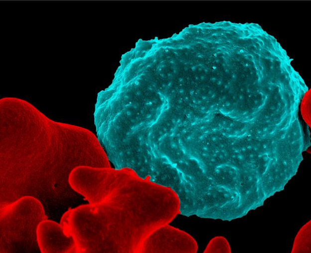
Caption: Colorized scanning electron micrograph of a blood cell infected with malaria parasites (blue with dots) surrounded by uninfected cells (red).
Credit: National Institute of Allergy and Infectious Diseases, NIH
As a volunteer physician in a small hospital in Nigeria 30 years ago, I was bitten by lots of mosquitoes and soon came down with headache, chills, fever, and muscle aches. It was malaria. Fortunately, the drug available to me then was effective, but I was pretty sick for a few days. Since that time, malarial drug resistance has become steadily more widespread. In fact, the treatment that cured me would be of little use today. Combination drug therapies including artemisinin have been introduced to take the place of the older drugs [1], but experts are concerned the mosquito-borne parasites that cause malaria are showing signs of drug resistance again.
So, researchers have been searching the genome of Plasmodium falciparum, the most-lethal species of the malaria parasite, for potentially better targets for drug or vaccine development. You wouldn’t think such work would be too tough because the genome of P. falciparum was sequenced more than 15 years ago [2]. Yet it’s proven to be a major challenge because the genetic blueprint of this protozoan parasite has an unusual bias towards two nucleotides (adenine and thymine), which makes it difficult to use standard research tools to study the functions of its genes.
Now, using a creative new spin on an old technique, an NIH-funded research team has solved this difficult problem and, for the first time, completely characterized the genes in the P. falciparum genome [3]. Their work identified 2,680 genes essential to P. falciparum’s growth and survival in red blood cells, where it does the most damage in humans. This gene list will serve as an important guide in the years ahead as researchers seek to identify the equivalent of a malarial Achilles heel, and use that to develop new and better ways to fight this deadly tropical disease.
Could Zika Virus Have Lasting Impact on Male Fertility?
Posted on by Dr. Francis Collins
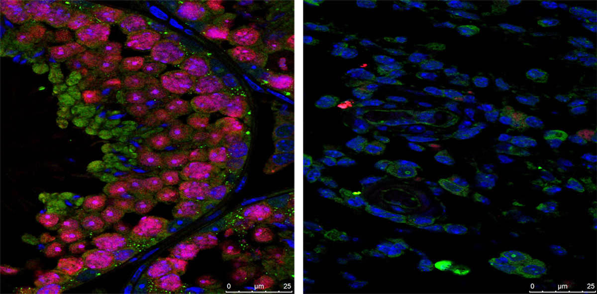
Caption: Immunofluorescence staining showing that the testes of Zika-free mice (left) are full of developing sperm (pink), while the testes of Zika-infected mice (right) contain very few sperm.
Credit: Prabagaran Esakky, Washington University School of Medicine, St. Louis
Recent research has shown that the mosquito-borne Zika virus has the potential to cause serious health problems, including severe birth defects in humans. But the damaging effects of Zika might not end there: results of a new mouse study show that the virus may also have an unexpected negative—and possibly long-lasting—impact on male fertility.
In work published in the journal Nature, an NIH-funded research team found that Zika infections can persist for many weeks in the reproductive systems of male mice [1]. As a result of this infection, levels of testosterone and other sex hormones drop, sperm counts fall, and, in some animals, the testicles shrink to 1/10th of their normal size, possibly irreversibly. All of this adds up to Zika-infected male mice that are significantly less fertile than their healthy counterparts—producing about a quarter as many viable offspring as normal when mated with female mice. While mice are certainly not humans, the results underscore the urgent need for additional research to examine the full spectrum of Zika’s health effects in men, women, and children of both sexes.
Zika Vaccine: Two Candidates Show Promise in Mice
Posted on by Dr. Francis Collins
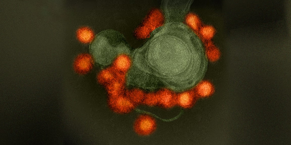
Caption: Zika virus (red), isolated from a microcephaly case in Brazil. The virus is associated with cellular membranes in the center.
Credit: NIAID
Last February, the World Health Organization declared a public health emergency over concerns about very serious birth defects in Brazil and their possible link to Zika virus. But even before then, concerns about the unprecedented spread of Zika virus in Brazil and elsewhere in Latin America had prompted NIH-funded scientists to step up their efforts to combat this emerging infectious disease threat. Over the last year, research aimed at understanding the mosquito-borne virus has progressed rapidly, and we now appear to be getting closer to a Zika vaccine.
In a recent study in the journal Nature, researchers found that a single dose of either of two experimental vaccines completely protected mice against a major viral strain responsible for the Zika outbreak in Brazil [1]. Caution is certainly warranted when extrapolating these (or any other) findings from mice to people. But, taking into account the fact that researchers have already developed safe and effective human vaccines for several related viruses, the new work represents a very encouraging milestone on the road toward a much-needed Zika vaccine for humans.
Snapshots of Life: Portrait of Zika Virus
Posted on by Dr. Francis Collins
Credit: David Goodsell, The Scripps Research Institute
This lively interplay of shape and color is an artistic rendering of the Zika virus preparing to enter a cell (blue) by binding to its protein receptors (green). The spherical structures (pink) represent two Zika viruses in a blood vessel filled with blood plasma cells (tan). The virus in the middle in cross section shows viral envelope proteins (red) studding the outer surface, with membrane proteins (pink) embedded in a fatty layer of lipids (light purples). In the innermost circle, you can see the viral genome (yellow) coiled around capsid proteins (orange).
This image was sketched and hand-painted with watercolors by David Goodsell, a researcher and illustrator at The Scripps Research Institute, La Jolla, CA. Goodsell put paint and science to paper as part of the “Molecule of the Month” series run by RCSB Protein Data Bank (PDB), which NIH co-supports with the National Science Foundation and the Department of Energy. The PDB, which contains structural data on thousands of proteins and small molecules, uses its “Molecule of the Month” series to help students visualize a molecule or virus and to encourage their exploration of structural biology and its applications to medicine.
Zika and Birth Defects: The Evidence Mounts
Posted on by Dr. Francis Collins

Caption: Human neural progenitor cells (gray) infected with Zika virus (green) increased the enzyme caspase-3 (red), suggesting increased cell death.
Credit: Sarah C. Ogden, Florida State University, Tallahassee
Recently, public health officials have raised major concerns over the disturbing spread of the mosquito-borne Zika virus among people living in and traveling to many parts of Central and South America [1]. While the symptoms of Zika infection are typically mild, grave concerns have arisen about its potential impact during pregnancy. The concerns stem from the unusual number of births of children with microcephaly, a very serious condition characterized by a small head and damaged brain, coinciding with the spread of Zika virus. Now, two new studies strengthen the connection between Zika and an array of birth defects, including, but not limited to, microcephaly.
In the first study, NIH-funded laboratory researchers show that Zika virus can infect and kill human neural progenitor cells [2]. Those progenitor cells give rise to the cerebral cortex, a portion of the brain often affected in children with microcephaly. The second study, involving a small cohort of women diagnosed with Zika virus during their pregnancies in Rio de Janeiro, Brazil, suggests that the attack rate is disturbingly high, and microcephaly is just one of many risks to the developing fetus. [3]
Gene Drive Research Takes Aim at Malaria
Posted on by Dr. Francis Collins
 Malaria has afflicted humans for millennia. Even today, the mosquito-borne, parasitic disease claims more than a half-million lives annually [1]. Now, in a study that has raised both hope and concern, researchers have taken aim at this ancient scourge by using one of modern science’s most powerful new technologies—the CRISPR/Cas9 gene-editing tool—to turn mosquitoes from dangerous malaria vectors into allies against infection [2].
The secret behind this new strategy is the “gene drive,” which involves engineering an organism’s genome in a way that intentionally spreads, or drives, a trait through its population much faster than is possible by normal Mendelian inheritance. The concept of gene drive has been around since the late 1960s [3]; but until the recent arrival of highly precise gene editing tools like CRISPR/Cas9, the approach was largely theoretical. In the new work, researchers inserted into a precise location in the mosquito chromosome, a recombinant DNA segment designed to block transmission of malaria parasites. Importantly, this segment also contained a gene drive designed to ensure the trait was inherited with extreme efficiency. And efficient it was! When the gene-drive engineered mosquitoes were mated with normal mosquitoes in the lab, they passed on the malaria-blocking trait to 99.5 percent of their offspring (as opposed to 50 percent for Mendelian inheritance).
Next Page