Zika clinical trial
Zika Vaccine: Two Candidates Show Promise in Mice
Posted on by Dr. Francis Collins
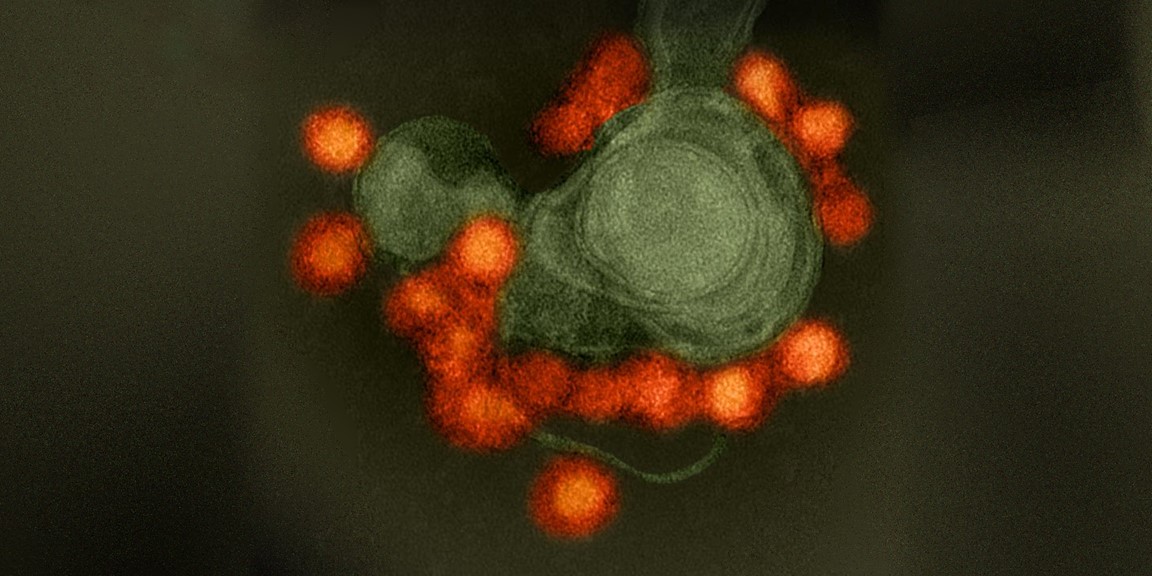
Caption: Zika virus (red), isolated from a microcephaly case in Brazil. The virus is associated with cellular membranes in the center.
Credit: NIAID
Last February, the World Health Organization declared a public health emergency over concerns about very serious birth defects in Brazil and their possible link to Zika virus. But even before then, concerns about the unprecedented spread of Zika virus in Brazil and elsewhere in Latin America had prompted NIH-funded scientists to step up their efforts to combat this emerging infectious disease threat. Over the last year, research aimed at understanding the mosquito-borne virus has progressed rapidly, and we now appear to be getting closer to a Zika vaccine.
In a recent study in the journal Nature, researchers found that a single dose of either of two experimental vaccines completely protected mice against a major viral strain responsible for the Zika outbreak in Brazil [1]. Caution is certainly warranted when extrapolating these (or any other) findings from mice to people. But, taking into account the fact that researchers have already developed safe and effective human vaccines for several related viruses, the new work represents a very encouraging milestone on the road toward a much-needed Zika vaccine for humans.
Snapshots of Life: Portrait of Zika Virus
Posted on by Dr. Francis Collins
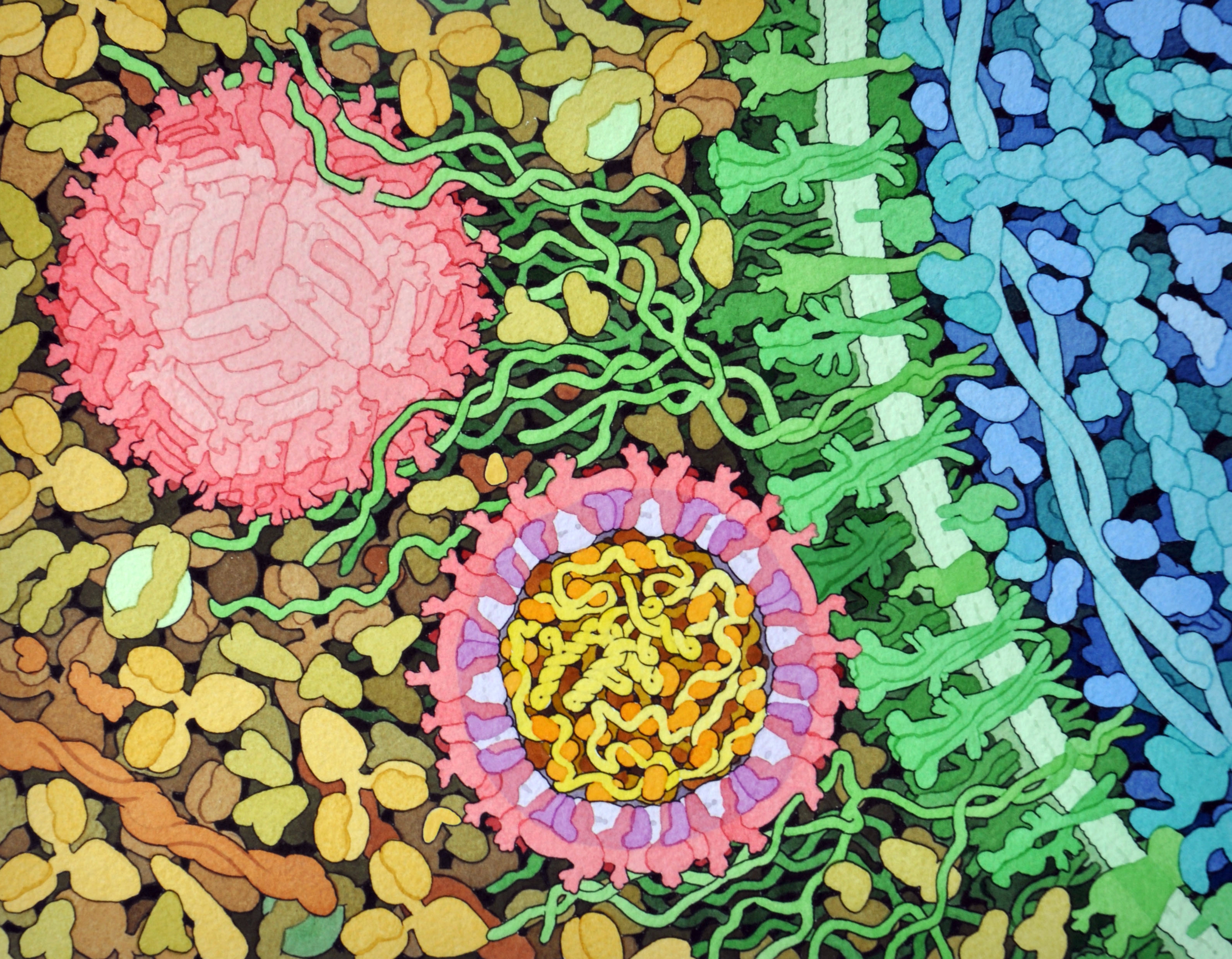
Credit: David Goodsell, The Scripps Research Institute
This lively interplay of shape and color is an artistic rendering of the Zika virus preparing to enter a cell (blue) by binding to its protein receptors (green). The spherical structures (pink) represent two Zika viruses in a blood vessel filled with blood plasma cells (tan). The virus in the middle in cross section shows viral envelope proteins (red) studding the outer surface, with membrane proteins (pink) embedded in a fatty layer of lipids (light purples). In the innermost circle, you can see the viral genome (yellow) coiled around capsid proteins (orange).
This image was sketched and hand-painted with watercolors by David Goodsell, a researcher and illustrator at The Scripps Research Institute, La Jolla, CA. Goodsell put paint and science to paper as part of the “Molecule of the Month” series run by RCSB Protein Data Bank (PDB), which NIH co-supports with the National Science Foundation and the Department of Energy. The PDB, which contains structural data on thousands of proteins and small molecules, uses its “Molecule of the Month” series to help students visualize a molecule or virus and to encourage their exploration of structural biology and its applications to medicine.