materials science
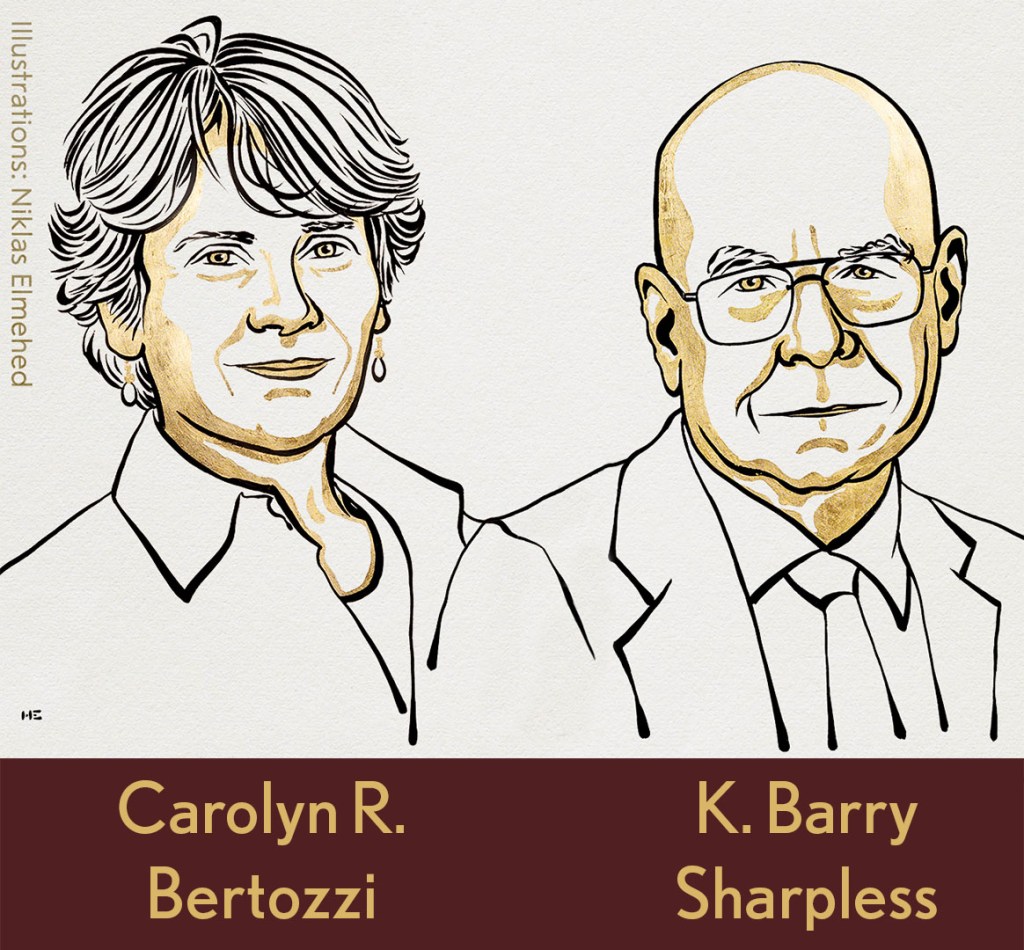
The Chemistry Clicked: Two NIH-Supported Researchers Win 2022 Nobel Prize in Chemistry
Posted on by Lawrence Tabak, D.D.S., Ph.D.
Through the years, NIH has supported a total of 169 researchers who have received or shared 101 Nobel Prizes. That’s quite a testament to the world-leading science that NIH pursues and its continued impact on improving human health and well-being.
Those numbers include the news late last week that the 2022 Nobel Prize in Chemistry was shared by two long-time grantees for their work on a transformative scientific approach known as “click chemistry.” This form of chemistry has made it possible for researchers to snap together, like LEGO pieces, molecular building blocks to form hybrid biomolecules, often with easy-to-track imaging agents attached. Not only has click chemistry expanded our ability to explore the molecular underpinnings of a wide range of biological processes, but it has provided us with new tools for developing drugs, diagnostics, and a wide array of “smart” materials.
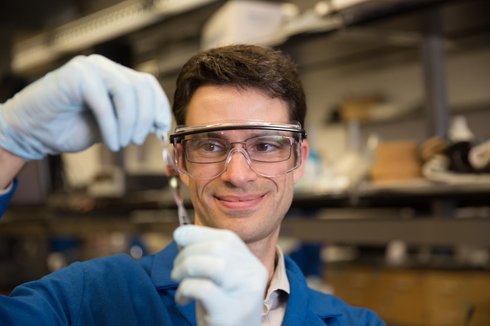
For K. Barry Sharpless, Scripps Research, La Jolla, CA, October 5, 2022 marked the second time that he’s received an early-morning congratulatory call from The Royal Swedish Academy of Sciences. The first such call came in 2001, when Sharpless got the news that he was a co-winner of the Nobel Prize in Chemistry for his discovery of asymmetric catalytic reactions.
This time around, Sharpless was recognized for his groundbreaking studies in the mid-1990s with click chemistry, a term that he coined himself. His initial work established click chemistry as a fast-and-reliable way to attach molecules of interest in the lab [1]. He and co-recipient Morten Meldal, University of Copenhagen, Denmark, who is not funded by NIH, then independently introduced a copper-catalyzed click that further refined the chemistry and helped popularize it across biology and the material sciences [2,3].
For Carolyn R. Bertozzi of Stanford University, Palo Alto, CA, it is her first Nobel. Bertozzi was recognized for expanding the use of click chemistry with so-called bioorthogonal chemistry, which is a copper-free version of the approach that can be used inside living cells without the risk of metal-associated toxicities [4,5].
Bertozzi’s work has been especially interesting to me because of her focus on glycans, which I’ve studied throughout my career. Glycans are the carbohydrate molecules that coat the surfaces of our cells and most secreted proteins. They are essential to life, and, in higher organisms, play fundamental roles in basic processes such as metabolism, immunity, and cellular communication.
Glycans also remain poorly understood, largely because, until recently, they have been so difficult for basic scientists to study with traditional techniques. That has changed with development of new tools to study glycans and the enzymes that assemble them. My long-time collaborator, Kelly Ten Hagen, a senior investigator at NIH’s National Institute of Dental and Craniofacial Research, and I collaborated with Carolyn on identifying small molecules that inhibit the enzyme responsible for the first step in mucin-type O-glycosylation [6]
In the early 2000s, Bertozzi and her team introduced bioorthogonal chemistry, which enabled researchers to label glycans and visualize them in a range of cells and living organisms. Her team’s pioneering approach quickly became an essential tool in basic science labs around the world that study glycans, leading to a number of stunning discoveries that would have otherwise been difficult or impossible.
For clinical researchers, click chemistry has emerged as a workhorse in drug discovery and the improved targeting of cancer chemotherapies and other small-molecule drugs. The approach also is being used to improve delivery of antibody-based therapies and to create new biomaterials. Meanwhile, in the material sciences, click chemistry has been used to solve a number of problems in working with polymers and to expand their industrial uses.
Click chemistry is an excellent example of how advances in basic science can build the foundation for a wide range of practical applications, including those aimed at improving human health. It also highlights the value of strong, sustained public funding for fundamental research, and NIH is proud to have supported Sharpless continuously since 1975 and Bertozzi since 1999. I send my sincere congratulations to both of these most-deserving scientists.
References:
[1] Click Chemistry: Diverse chemical function from a few good reactions. Kolb, HC, Finn, MG, Sharpless, KB. Angew. Chem. Int. Ed. 2001, 40 (11), 2004–2021
[2] A stepwise huisgen cycloaddition process: Copper(I)-catalyzed regioselective “Llgation” of azides and terminal alkynes. Rostovtsev VV, Green LG, Fokin VV, Sharpless KB. Angew. Chem. Int. Ed. 2002, 41 (14), 2596–2599.
[3] Peptidotriazoles on solid phase: [1,2,3]-Triazoles by regiospecific copper(I)-catalyzed 1,3-dipolar cycloadditions of terminal alkynes to azides. Tornøe CW, Sengeløv H, Meldal M. J. Org. Chem. 2002, 67 (9), 3057–3064.
[4] A strain-promoted [3 + 2] azide−alkyne cycloaddition for covalent modification of biomolecules in living systems. Agard NJ, Prescher JA, Bertozzi CR. J. Am. Chem. Soc. 2004, 126 (46), 15046–15047
[5] In vivo imaging of membrane associated glycans in developing zebrafish. Laughlin ST, Baskin JM, Amacher SL, Bertozzi CR. Science 2008, 320 (5876), 664–667.
[6] Small molecule inhibitors of mucin-type O-glycosylation from a uridine-based library. Hang, HC, Yu, C, Ten Hagen, KG, Tian, E, Winans, KA, Tabak, LA, Bertozzi, Chem Biol. 2004 Jul;11(7):1009-1016.
Links:
The Nobel Prize in Chemistry 2022 (The Royal Swedish Academy of Sciences, Stockholm)
Video: Announcement of the 2022 Nobel Prize in Chemistry (YouTube)
Click Chemistry and Bioorthogonal Chemistry (The Royal Swedish Academy of Sciences)
Sharpless Lab (Scripps Research, La Jolla, CA)
Bertozzi Group (Stanford University, Palo Alto, CA)
NIH Support:
K. Barry Sharpless: National Institute of General Medical Sciences
Carolyn R. Bertozzi: National Cancer Institute; National Institute of Allergy and Infectious Diseases; National Institute of General Medical Sciences
Tackling Fibrosis with Synthetic Materials
Posted on by Dr. Francis Collins

When injury strikes a limb or an organ, our bodies usually heal quickly and correctly. But for some people, the healing process doesn’t shut down properly, leading to excess fibrous tissue, scarring, and potentially life-threatening organ damage.
This permanent scarring, known as fibrosis, can occur in almost every tissue of the body, including the heart and lungs. With support from a 2019 NIH Director’s New Innovator Award, April Kloxin is applying her expertise in materials science and bioengineering to build sophisticated fibrosis-in-a-dish models for unraveling this complex process in her lab at the University of Delaware, Newark.
Though Kloxin is interested in all forms of fibrosis, she’s focusing first on the incurable and often-fatal lung condition called idiopathic pulmonary fibrosis (IPF). This condition, characterized by largely unexplained thickening and stiffening of lung tissue, is diagnosed in about 50,000 people each year in the United States.
IPF remains poorly understood, in part because it often is diagnosed when the disease is already well advanced. Kloxin hopes to turn back the clock and start to understand the disease at an earlier stage, when interventions might be more successful. The key is to develop a model that better recapitulates the complexity and irreversibility of the disease process in people.
Building that better model starts with simulating the meshwork of collagen and other proteins in the extracellular matrix (ECM) that undergird every tissue and organ in the body. The ECM’s interactions with our cells are essential in wound healing and, when things go wrong, also in causing fibrosis.
Kloxin will build three-dimensional hydrogels, crosslinked sponge-like networks of polymers, peptides, and proteins, with structures that more accurately capture the biological complexities of human tissues, including the ECMs within fibrous collagen-rich microenvironments. Her synthetic matrices can be triggered with light to lock in place and stiffen. The matrices also will make it possible to culture the lung’s epithelium, or outermost layer of cells, and connective tissue that surrounds it, to study cellular responses as the model shifts from a healthy and flexible to a stiffened, disease-like state.
Kloxin and her team will also integrate into their model system lung cells that have been engineered to fluoresce or light up under a microscope when the wound-healing program activates. Such fluorescent reporters will allow her team to watch for the first time how different cells and their nearby microenvironment respond as the composition of the ECM changes and stiffens. With this system, she’ll also be able to search for small molecules with the ability to turn off excessive wound healing.
The hope is that what’s learned with her New Innovator Award will lead to fresh insights and ultimately new treatments for this mysterious, hard-to-treat condition. But the benefits could be even more wide-ranging. Kloxin thinks that her findings will have implications for the prevention and treatment of other fibrotic diseases as well.
Links:
Idiopathic Pulmonary Fibrosis (National Heart, Lung, and Blood Institute/NIH)
April Kloxin Group (University of Delaware, Newark)
Kloxin Project Information (NIH RePORTER)
NIH Director’s New Innovator Award (Common Fund)
NIH Support: Common Fund; National Heart, Lung, and Blood Institute
Taking Microfluidics to New Lengths
Posted on by Dr. Francis Collins
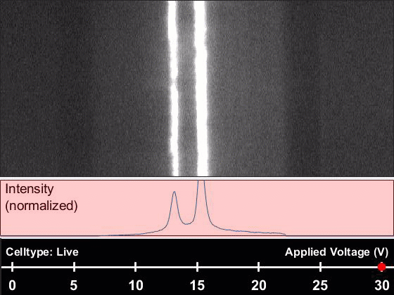
Caption: Microfluidic fiber sorting a solution containing either live or dead cells. The type of cell being imaged and the real time voltage (30v) is displayed at bottom. It is easy to imagine how this could be used to sort a mixture of live and dead cells. Credit: Yuan et al., PNAS
Microfluidics—the manipulation of fluids on a microscopic scale— has made it possible to produce “lab-on-a-chip” devices that detect, for instance, the presence of Ebola virus in a single drop of blood. Now, researchers hope to apply the precision of microfluidics to a much broader range of biomedical problems. Their secret? Move the microlab from chips to fibers.
To do this, an NIH-funded team builds microscopic channels into individual synthetic polymer fibers reaching 525 feet, or nearly two football fields long! As shown in this video, the team has already used such fibers to sort live cells from dead ones about 100 times faster than current methods, relying only on natural differences in the cells’ electrical properties. With further design and development, the new, fiber-based systems hold great promise for, among other things, improving kidney dialysis and detecting metastatic cancer cells in a patient’s bloodstream.
Snapshots of Life: Virus Hunting with Carbon Nanotubes
Posted on by Dr. Francis Collins

Credit: Penn State University
The purple pods that you see in this scanning electron micrograph are the H5N2 avian flu virus, a costly threat to the poultry and egg industry and, in very rare instances, a health risk for humans. However, these particular pods are unlikely to infect anything because they are trapped in a gray mesh of carbon nanotubes. Made by linking carbon atoms into a cylindrical pattern, such nanotubes are about 10,000 times smaller than width of a human hair.
The nanotubes above have been carefully aligned on a special type of silicon chip called a carbon-nanotube size-tunable-enrichment-microdevice (CNT-STEM). As described recently in Science Advances, this ultrasensitive device is designed to capture viruses rapidly based on their size, not their molecular characteristics [1]. This unique feature enables researchers to detect completely unknown viruses, even when they are present in extremely low numbers. In proof-of-principle studies, CNT-STEM made it possible to collect and detect viruses in a sample at concentrations 100 times lower than with other methods, suggesting the device and its new approach will be helpful in the ongoing hunt for new and emerging viruses, including those that infect people.
Precision Oncology: Nanoparticles Target Bone Cancers in Dogs
Posted on by Dr. Francis Collins
Credit: L. Brian Stauffer
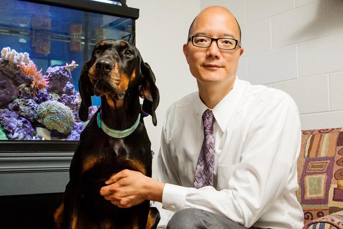
Many people share their homes with their pet dogs. Spending years under the same roof with the same environmental exposures, people and dogs have something else in common that sometimes gets overlooked. They can share some of the same diseases, such as diabetes and cancer. By studying these diseases in dogs, researchers can learn not only to improve care for people but for their canine friends as well.
As a case in point, an NIH-funded team of researchers recently tested a new method of delivering chemotherapy drugs for osteosarcoma, a bone cancer that affects dogs and people, typically teenagers and older adults. Their studies in dogs undergoing treatment for osteosarcoma suggest that specially engineered, bone-seeking nanoparticles might safely deliver anti-cancer drugs precisely to the places where they are most needed. These early findings come as encouraging news for the targeted treatment of inoperable bone cancers and other malignancies that spread to bone.
Snapshots of Life: Finding a Cube for Cancer
Posted on by Dr. Francis Collins
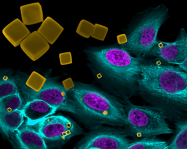
Jenolyn F. Alexander and Biana Godin, Houston Methodist Research Institute; Veronika Kozlovskaya and Eugenia Kharlampieva, University of Alabama at Birmingham.
Creative photographers have long experimented with superimposing images, one over the other, to produce striking visual effects. Now a group of NIH-supported scientists at Houston Methodist Research Institute and their colleagues have done the same thing to highlight their work in the emerging field of cancer nanomedicine, using microscopic materials to deliver cancer treatments with potentially greater precision. In the process, the researchers generated a photographic work of art that was a winner in the Federation of American Societies for Experimental Biology 2015 Bioart competition.
The gold cubes are man-made polymer microcarriers, just 2 micrometers wide (by comparison, human cells generally range in diameter from 7 to 20 micrometers), designed to transport chemotherapy drugs directly to tumor cells. These experimental cubes, enlarged in the upper left part of the photo with a scanning electron microscope for better viewing, have been superimposed onto a second photograph snapped with a confocal fluorescence microscope. It shows similar cube-shaped microcarriers (yellow) inside cultured breast cancer cells (nucleus is purple, cytoplasm is turquoise).
Creative Minds: Stretching the Limits of Wearable Devices
Posted on by Dr. Francis Collins
Darren Lipomi/ Credit: UC, San Diego
Whether it’s a pedometer dangling from a belt loop or a skin patch to monitor heart rate and hydration levels, wearable and mobile devices have become essential gear for many of today’s fitness minded. But Darren Lipomi, a nanoengineer at the University of California, San Diego, envisions even more impressive things to come for optimizing workouts and bringing greater precision to health care. Lipomi is helping to build a future of “stretchable electronics,” semiconducting devices that will more seamlessly integrate with the contours of our bodies, outside and even inside, to monitor vital signs, muscle activity, metabolic changes, and organ function—to name just a few possibilities.
Lipomi and his colleagues specifically want to create a new class of semiconducting polymer that has the mechanical properties of human skin. This transparent “electronic skin” will have a soft elasticity to conform to shape, sense contact, absorb blunt force, and even self heal when dinged. It will do all of this—and possibly more—while continuously and wirelessly performing its programmed health-monitoring function. To help Lipomi build this future of real-time health monitoring, he has been awarded a 2015 NIH Director’s New Innovator Award. This NIH award supports exceptionally creative new investigators who propose highly innovative projects with the potential for unusually high impact.