confocal fluorescence microscope
Snapshots of Life: Finding a Cube for Cancer
Posted on by Dr. Francis Collins
Jenolyn F. Alexander and Biana Godin, Houston Methodist Research Institute; Veronika Kozlovskaya and Eugenia Kharlampieva, University of Alabama at Birmingham.
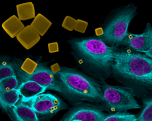
Creative photographers have long experimented with superimposing images, one over the other, to produce striking visual effects. Now a group of NIH-supported scientists at Houston Methodist Research Institute and their colleagues have done the same thing to highlight their work in the emerging field of cancer nanomedicine, using microscopic materials to deliver cancer treatments with potentially greater precision. In the process, the researchers generated a photographic work of art that was a winner in the Federation of American Societies for Experimental Biology 2015 Bioart competition.
The gold cubes are man-made polymer microcarriers, just 2 micrometers wide (by comparison, human cells generally range in diameter from 7 to 20 micrometers), designed to transport chemotherapy drugs directly to tumor cells. These experimental cubes, enlarged in the upper left part of the photo with a scanning electron microscope for better viewing, have been superimposed onto a second photograph snapped with a confocal fluorescence microscope. It shows similar cube-shaped microcarriers (yellow) inside cultured breast cancer cells (nucleus is purple, cytoplasm is turquoise).