MERS
Finding New Ways to Fight Coronavirus … From Studying Bats
Posted on by Dr. Francis Collins

David Veesler has spent nearly 20 years imaging in near-atomic detail the parts of various viruses, including coronaviruses, that enable them to infect Homo sapiens. In fact, his lab at the University of Washington, Seattle, was the first to elucidate the 3D architecture of the now infamous spike protein, which coronaviruses use to gain entry into human cells [1]. He uses these fundamental insights to guide the design of vaccines and therapeutics, including promising monoclonal antibodies.
Now, Veesler and his lab are turning to another mammal in their search for new leads for the next generation of antiviral treatments, including ones aimed at the coronavirus that causes COVID-19, SARS-CoV-2. With support from a 2020 NIH Director’s Pioneer Award, Veesler will study members of the order Chiroptera. Or, more colloquially, bats.
Why bats? Veesler says bats are remarkable creatures. They are the only mammals capable of sustained flight. They rarely get cancer and live unusually long lives for such small creatures. More importantly for Veesler’s research, bats host a wide range of viruses—more than any other mammal species. Despite carrying all of these viruses, bats rarely show symptoms of being sick. Yet they are the source for many of the viruses that have spilled over into humans with devastating effect, including rabies, Ebola virus, Nipah and Hendra viruses, severe acute respiratory syndrome coronavirus (SARS-CoV), and, likely, SARS-CoV-2.
Beyond what is already known about bats’ intriguing qualities, Veesler says humans still have much to discover about these flying mammals, including how their immune systems cope with such an onslaught of viral invaders. For example, it turns out that a bat’s learned, or adaptive, immune system is, for the most part, uncharted territory. As such, it offers an untapped source of potentially promising viral inhibitors just waiting to be unearthed, fully characterized, and then used to guide the development of new kinds of anti-viral therapeutics.
In his studies, Veesler will work with collaborators studying bats around the world to characterize their antibody production. He wants to learn how these antibodies contribute to bats’ impressive ability to tolerate viruses and other pathogens. What is it about the structure of bat antibodies that make them different from human antibodies? And, how can those structural differences serve as blueprints for promising new treatments to combat many potentially deadly viruses?
Interestingly, Veesler’s original grant proposal makes no mention of SARS-CoV-2 or COVID-19. That’s because he submitted it just months before the first reports of the novel coronavirus in Wuhan, China. But Veesler doesn’t consider himself a visionary by expanding his research to bats. He and others had been working on closely related coronaviruses for years, inspired by earlier outbreaks, including SARS in 2002 and Middle East respiratory syndrome (MERS) in 2012 (although MERS apparently came from camels). The researcher didn’t see SARS-CoV-2 coming, but he recognized the potential for some kind of novel coronavirus outbreak in the future.
These days, the Veesler lab has been hard at work to understand SARS-CoV-2 and the human immune response to the virus. His team showed that SARS-CoV-2 uses the human receptor ACE2 to gain entry into our cells [2]. He’s also a member of the international research team that identified a human antibody, called S309, from a person who’d been infected with SARS in 2003. This antibody is showing promise for treating COVID-19 [3], now in a phase 3 clinical trial in the United States.
In another recent study, reported as a pre-print in bioRxiv, Veesler’s team mapped dozens of distinct human antibodies capable of neutralizing SARS-CoV-2 by their ability to hit viral targets outside of the well-known spike protein [4]. Such discoveries may form the basis for new and promising combinations of antibodies to treat COVID-19 that won’t be disabled by concerning new variations in the SARS-CoV-2 spike protein. Perhaps, in the future, such therapeutic cocktails may include modified bat-inspired antibodies too.
References:
[1] Cryo-electron microscopy structure of a coronavirus spike glycoprotein trimer. Walls AC, Tortorici MA, Bosch BJ, Frenz B, Rottier PJM, DiMaio F, Rey FA, Veesler D. Nature. 2016 Mar 3;531(7592):114-117.
[2] Structure, function, and antigenicity of the SARS-CoV-2 spike glycoprotein. Walls AC, Park YJ, Tortorici MA, Wall A, McGuire AT, Veesler D. Cell. 2020 Apr 16;181(2):281-292.e6.
[3] Cross-neutralization of SARS-CoV-2 by a human monoclonal SARS-CoV antibody. Pinto D, Park YJ, Beltramello M, Veesler D, Cortil D, et al. Nature.18 May 2020 [Epub ahead of print]
[4] N-terminal domain antigenic mapping reveals a site of vulnerability for SARS-CoV-2. McCallum M, Marco A, Lempp F, Tortorici MA, Pinto D, Walls AC, Whelan SPJ, Virgin HW, Corti D, Pizzuto MS, Veesler D, et al. bioRxiv. 2021 Jan 14.
Links:
COVID-19 Research (NIH)
Veesler Lab (University of Washington, Seattle)
Veesler Project Information (NIH RePORTER)
NIH Director’s Pioneer Award Program (Common Fund)
NIH Support: Common Fund; National Institute of Allergy and Infectious Diseases
Celebrating the Gift of COVID-19 Vaccines
Posted on by Dr. Francis Collins

The winter holidays are traditionally a time of gift-giving. As fatiguing as 2020 and the COVID-19 pandemic have been, science has stepped up this year to provide humankind with a pair of truly hopeful gifts: the first two COVID-19 vaccines.
Two weeks ago, the U.S. Food and Drug Administration (FDA) granted emergency use authorization (EUA) to a COVID-19 vaccine from Pfizer/BioNTech, enabling distribution to begin to certain high-risk groups just three days later. More recently, the FDA granted an EUA to a COVID-19 vaccine from the biotechnology company Moderna, Cambridge, MA. This messenger RNA (mRNA) vaccine, which is part of a new approach to vaccination, was co-developed by NIH’s National Institute of Allergy and Infectious Diseases (NIAID). The EUA is based on data showing the vaccine is safe and 94.5 percent effective at protecting people from infection with SARS-CoV-2, the coronavirus that causes COVID-19.
Those data on the Moderna vaccine come from a clinical trial of 30,000 individuals, who generously participated to help others. We can’t thank those trial participants enough for this gift. The distribution of millions of Moderna vaccine doses is expected to begin this week.
It’s hard to put into words just how remarkable these accomplishments are in the history of science. A vaccine development process that used to take many years, often decades, has been condensed to about 11 months. Just last January, researchers started out with a previously unknown virus and we now have not just one, but two, vaccines that will be administered to millions of Americans before year’s end. And the accomplishments don’t end there—several other types of COVID-19 vaccines are also on the way.
It’s important to recognize that this couldn’t have happened without the efforts of many scientists working tirelessly behind the scenes for many years prior to the pandemic. Among those who deserve tremendous credit are Kizzmekia Corbett, Barney Graham, John Mascola, and other members of the amazing team at the Dale and Betty Bumpers Vaccine Research Center at NIH’s National Institute of Allergy and Infectious Diseases (NIAID).
When word of SARS-CoV-2 emerged, Corbett, Graham, and other NIAID researchers had already been studying other coronaviruses for years, including those responsible for earlier outbreaks of respiratory disease. So, when word came that this was a new coronavirus outbreak, they were ready to take action. It helped that they had paid special attention to the spike proteins on the surface of coronaviruses, which have turned out to be the main focus the COVID-19 vaccines now under development.
The two vaccines currently authorized for administration in the United States work in a unique way. Their centerpiece is a small, non-infectious snippet of mRNA. Our cells constantly produce thousands of mRNAs, which provide the instructions needed to make proteins. When someone receives an mRNA vaccine for COVID-19, it tells the person’s own cells to make the SARS-CoV-2 spike protein. The person’s immune system then recognizes the viral spike protein as foreign and produces antibodies to eliminate it.
This vaccine-spurred encounter trains the human immune system to remember the spike protein. So, if an actual SARS-CoV-2 virus tries to infect a vaccinated person weeks or months later, his or her immune system will be ready to fend it off. To produce the most vigorous and durable immunity against the virus, people will need to get two shots of mRNA vaccine, which are spaced several weeks to a month apart, depending on the vaccine.
Some have raised concerns on social media that mRNA vaccines might alter the DNA genome of someone being vaccinated. But that’s not possible, since this mRNA doesn’t enter the nucleus of the cell where DNA is located. Instead, the vaccine mRNAs stay in the outer part of the cell (the cytoplasm). What’s more, after being transcribed into protein just one time, the mRNA quickly degrades. Others have expressed concerns about whether the vaccine could cause COVID-19. That is not a risk because there’s no whole virus involved, just the coding instructions for the non-infectious spike protein.
An important advantage of mRNA is that it’s easy for researchers to synthesize once they know the nucleic acid sequence of a target viral protein. So, the gift of mRNA vaccines is one that will surely keep on giving. This new technology can now be used to speed the development of future vaccines. After the emergence of the disease-causing SARS, MERS, and now SARS-CoV-2 viruses, it would not be surprising if there are other coronavirus health threats in our future. Corbett and her colleagues are hoping to design a universal vaccine that can battle all of them. In addition, mRNA vaccines may prove effective for fighting future pandemics caused by other infectious agents and for preventing many other conditions, such as cancer and HIV.
Though vaccines are unquestionably our best hope for getting past the COVID-19 pandemic, public surveys indicate that some people are uneasy about accepting this disease-preventing gift. Some have even indicated they will refuse to take the vaccine. Healthy skepticism is a good thing, but decisions like this ought to be based on weighing the evidence of benefit versus risk. The results of the Pfizer and Moderna trials, all released for complete public scrutiny, indicate the potential benefits are high and the risks, low. Despite the impressive speed at which the new COVID-19 vaccines were developed, they have undergone and continue to undergo a rigorous process to generate all the data needed by the FDA to determine their long-term safety and effectiveness.
Unfortunately, the gift of COVID-19 vaccines comes too late for the more than 313,000 Americans who have died from complications of COVID-19, and many others who’ve had their lives disrupted and may have to contend with long-term health consequences related to COVID-19. The vaccines did arrive in record time, but all of us wish they could somehow have arrived even sooner to avert such widespread suffering and heartbreak.
It will be many months before all Americans who are willing to get a vaccine can be immunized. We need 75-80 percent of Americans to receive vaccines in order to attain the so-called “herd immunity” needed to drive SARS-CoV-2 away and allow us all to get back to a semblance of normal life.
Meanwhile, we all have a responsibility to do everything possible to block the ongoing transmission of this dangerous virus. Each of us needs to follow the three W’s: Wear a mask, Watch your distance, Wash your hands often.
When your chance for immunization comes, please roll up your sleeve and accept the potentially life-saving gift of a COVID-19 vaccine. In fact, I just got my first shot of the Moderna vaccine today along with NIAID Director Anthony Fauci, HHS Secretary Alex Azar, and some front-line healthcare workers at the NIH Clinical Center. Accepting this gift is our best chance to put this pandemic behind us, as we look forward to a better new year.
Links:
Coronavirus (COVID-19) (NIH)
Combat COVID (U.S. Department of Health and Human Services, Washington, D.C.)
Dale and Betty Bumpers Vaccine Research Center (National Institute of Allergy and Infectious Diseases/NIH)
Moderna (Cambridge, MA)
Pfizer (New York, NY)
BioNTech (Mainz, Germany)
Protein Mapping Study Reveals Valuable Clues for COVID-19 Drug Development
Posted on by Dr. Francis Collins
One way to fight COVID-19 is with drugs that directly target SARS-CoV-2, the novel coronavirus that causes the disease. That’s the strategy employed by remdesivir, the only antiviral drug currently authorized by the U.S. Food and Drug Administration to treat COVID-19. Another promising strategy is drugs that target the proteins within human cells that the virus needs to infect, multiply, and spread.
With the aim of developing such protein-targeted antiviral drugs, a large, international team of researchers, funded in part by the NIH, has precisely and exhaustively mapped all of the interactions that take place between SARS-CoV-2 proteins and the human proteins found within infected host cells. They did the same for the related coronaviruses: SARS-CoV-1, the virus responsible for outbreaks of Severe Acute Respiratory Syndrome (SARS), which ended in 2004; and MERS-CoV, the virus that causes the now-rare Middle East Respiratory Syndrome (MERS).
The goal, as reported in the journal Science, was to use these protein “interactomes” to uncover vulnerabilities shared by all three coronaviruses. The hope is that the newfound knowledge about these shared proteins—and the pathways to which they belong—will inform efforts to develop new kinds of broad-spectrum antiviral therapeutics for use in the current and future coronavirus outbreaks.
Facilitated by the Quantitative Biosciences Institute Research Group, the team, which included David E. Gordon and Nevan Krogan, University of California, San Francisco, and hundreds of other scientists from around the world, successfully mapped nearly 400 protein-protein interactions between SARS-CoV-2 and human proteins.
You can see one of these interactions in the video above. The video starts out with an image of the Orf9b protein of SARS-CoV-2, which normally consists of two linked molecules (blue and orange). But researchers discovered that Orf9b dissociates into a single molecule (orange) when it interacts with the human protein TOM70 (teal). Through detailed structural analysis using cryo-electron microscopy (cryo-EM), the team went on to predict that this interaction may disrupt a key interaction between TOM70 and another human protein called HSP90.
While further study is needed to understand all the details and their implications, it suggests that this interaction may alter important aspects of the human immune response, including blocking interferon signals that are crucial for sounding the alarm to prevent serious illness. While there is no drug immediately available to target Orf9b or TOM70, the findings point to this interaction as a potentially valuable target for treating COVID-19 and other diseases caused by coronaviruses.
This is just one intriguing example out of 389 interactions between SARS-CoV-2 and human proteins uncovered in the new study. The researchers also identified 366 interactions between human and SARS-CoV-1 proteins and 296 for MERS-CoV. They were especially interested in shared interactions that take place between certain human proteins and the corresponding proteins in all three coronaviruses.
To learn more about the significance of these protein-protein interactions, the researchers conducted a series of studies to find out how disrupting each of the human proteins influences SARS-CoV-2’s ability to infect human cells. These studies narrowed the list to 73 human proteins that the virus depends on to replicate.
Among them were the receptor for an inflammatory signaling molecule called IL-17, which has been suggested as an indicator of COVID-19 severity. Two other human proteins—PGES-2 and SIGMAR1—were of particular interest because they are targets of existing drugs, including the anti-inflammatory indomethacin for PGES-2 and antipsychotics like haloperidol for SIGMAR1.
To connect the molecular-level data to existing clinical information for people with COVID-19, the researchers looked to medical billing data for nearly 740,000 Americans treated for COVID-19. They then zeroed in on those individuals who also happened to have been treated with drugs targeting PGES-2 or SIGMAR1. And the results were quite striking.
They found that COVID-19 patients taking indomethacin were less likely than those taking an anti-inflammatory that doesn’t target PGES-2 to require treatment at a hospital. Similarly, COVID-19 patients taking antipsychotic drugs like haloperidol that target SIGMAR1 were half as likely as those taking other types of antipsychotic drugs to require mechanical ventilation.
More research is needed before we can think of testing these or similar drugs against COVID-19 in human clinical trials. Yet these findings provide a remarkable demonstration of how basic molecular and structural biological findings can be combined with clinical data to yield valuable new clues for treating COVID-19 and other viral illnesses, perhaps by repurposing existing drugs. Not only is NIH-supported basic science essential for addressing the challenges of the current pandemic, it is building a strong foundation of fundamental knowledge that will make us better prepared to deal with infectious disease threats in the future.
Reference:
[1] Comparative host-coronavirus protein interaction networks reveal pan-viral disease mechanisms. Gordon DE et al. Science. 2020 Oct 15:eabe9403.
Links:
Coronavirus (COVID-19) (NIH)
Krogan Lab (University of California, San Francisco)
NIH Support: National Institute of Allergy and Infectious Diseases; National Institute of Neurological Disorders and Stroke; National Institute of General Medical Sciences
Study Finds People Have Short-Lived Immunity to Seasonal Coronaviruses
Posted on by Dr. Francis Collins

A key metric in seeking to end the COVID-19 pandemic is the likely duration of acquired immunity, which is how long people infected with SARS-CoV-2, the novel coronavirus that causes COVID-19, are protected against reinfection. The hope is that acquired immunity from natural infection—or from vaccines—will be long-lasting, but data to confirm that’s indeed the case won’t be in for many months or years.
In the meantime, a useful place to look for clues is in long-term data on reinfections with other seasonal coronaviruses. Could the behavior of less life-threatening members of the coronavirus family give us some insight into what to expect from SARS-CoV-2?
A new study, published in the journal Nature Medicine, has taken exactly this approach. The researchers examined blood samples collected continuously from 10 healthy individuals since the 1980s for evidence of infections—and reinfections—with four common coronaviruses. Unfortunately, it’s not particularly encouraging news. The new data show that immunity to other coronaviruses tends to be short-lived, with reinfections happening quite often about 12 months later and, in some cases, even sooner.
Prior to the discovery of SARS-CoV-2, six coronaviruses were known to infect humans. Four are responsible for relatively benign respiratory illnesses that regularly circulate to cause the condition we recognize as the common cold. The other two are more dangerous and, fortunately, less common: SARS-CoV-1, the virus responsible for outbreaks of Severe Acute Respiratory Syndrome (SARS), which ended in 2004; and MERS-CoV, the virus that causes the now rare Middle East Respiratory Syndrome (MERS).
In the new study, a team led by Lia van der Hoek, University of Amsterdam, the Netherlands, set out to get a handle on reinfections with the four common coronaviruses: HCoV-NL63, HCoV-229E, HCoV-OC43, and HCoV-HKU1. This task isn’t as straightforward as it might sound. That’s because, like SARS-CoV-2, infections with such viruses don’t always produce symptoms that are easily tracked. So, the researchers looked instead to blood samples from 10 healthy individuals enrolled for decades in the Amsterdam Cohort Studies on HIV-1 Infection and AIDS.
To detect coronavirus reinfections, they measured increases in antibodies to a particular portion of the nucleocapsid of each coronavirus. The nucleocapsid is a protein shell that encapsulates a coronavirus’ genetic material and serves as important targets for antibodies. An increase in antibodies targeting the nucleocapsid indicated that a person was fighting a new infection with one of the four coronaviruses.
All told, the researchers examined a total of 513 blood samples collected at regular intervals—every 3 to 6 months. In those samples, the team’s analyses uncovered 3 to 17 coronavirus infections per study participant over more than 35 years. Reinfections occurred every 6 to 105 months. But reinfections happened most frequently about a year after a previous infection.
Not surprisingly, they also found that blood samples collected in the Netherlands during the summer months—June, July, August, and September—had the lowest rate of infections for all four seasonal coronaviruses, indicating a higher frequency of infections in winter in temperate countries. While it remains to be seen, it’s possible that SARS-CoV-2 ultimately may share the same seasonal pattern after the pandemic.
These findings show that annual reinfections are a common occurrence for all other common coronaviruses. That’s consistent with evidence that antibodies against SARS-CoV-2 decrease within two months of infection [2]. It also suggests that similar patterns of reinfection may emerge for SARS-CoV-2 in the coming months and years.
At least three caveats ought to be kept in mind when interpreting these data. First, the researchers tracked antibody levels but didn’t have access to information about actual illness. It’s possible that a rise in antibodies to a particular coronavirus might have provided exactly the response needed to convert a significant respiratory illness to a mild case of the sniffles or no illness at all.
Second, sustained immunity to viruses will always be disrupted if the virus is undergoing mutational changes and presenting a new set of antigens to the host; the degree to which that might have contributed to reinfections is not known. And, third, the role of cell-based immunity in fighting off coronavirus infections is likely to be significant, but wasn’t studied in this retrospective analysis.
To prepare for COVID-19 this winter, it’s essential to understand how likely a person who has recovered from the illness will be re-infected and potentially spread the virus to other people. While much more study is needed, the evidence suggests it will be prudent to proceed carefully and with caution when it comes to long-term immunity, whether achieved through naturally acquired infections or vaccination.
While we await a COVID-19 vaccine, the best way to protect yourself, your family, and your community is to take simple steps all of us can do today: maintain social distancing, wear a mask, avoid crowded indoor gatherings, and wash your hands.
References:
[1] Seasonal coronavirus protective immunity is short-lasting. Edridge AWD, Kaczorowska J, Hoste ACR, Bakker M, Klein M, Loens K, Jebbink MF, Matser A, Kinsella CM, Rueda P, Ieven M, Goossens H, Prins M, Sastre P, Deijs M, van der Hoek L. Nat Med. 2020 Sep 14. doi: 10.1038/s41591-020-1083-1. [Published online ahead of print.]
[2] Rapid decay of anti-SARS-CoV-2 antibodies in persons with mild Covid-19. Ibarrondo FJ, Fulcher JA, Goodman-Meza D, Elliott J, Hofmann C, Hausner MA, Ferbas KG, Tobin NH, Aldrovandi GM, Yang OO. N Engl J Med. 2020 Sep 10;383(11):1085-1087.
Links:
Coronavirus (COVID-19) (NIH)
Lia van der hoek (University of Amsterdam, the Netherlands)
Immune T Cells May Offer Lasting Protection Against COVID-19
Posted on by Dr. Francis Collins

Much of the study on the immune response to SARS-CoV-2, the novel coronavirus that causes COVID-19, has focused on the production of antibodies. But, in fact, immune cells known as memory T cells also play an important role in the ability of our immune systems to protect us against many viral infections, including—it now appears—COVID-19.
An intriguing new study of these memory T cells suggests they might protect some people newly infected with SARS-CoV-2 by remembering past encounters with other human coronaviruses. This might potentially explain why some people seem to fend off the virus and may be less susceptible to becoming severely ill with COVID-19.
The findings, reported in the journal Nature, come from the lab of Antonio Bertoletti at the Duke-NUS Medical School in Singapore [1]. Bertoletti is an expert in viral infections, particularly hepatitis B. But, like so many researchers around the world, his team has shifted their focus recently to help fight the COVID-19 pandemic.
Bertoletti’s team recognized that many factors could help to explain how a single virus can cause respiratory, circulatory, and other symptoms that vary widely in their nature and severity—as we’ve witnessed in this pandemic. One of those potential factors is prior immunity to other, closely related viruses.
SARS-CoV-2 belongs to a large family of coronaviruses, six of which were previously known to infect humans. Four of them are responsible for the common cold. The other two are more dangerous: SARS-CoV-1, the virus responsible for the outbreak of Severe Acute Respiratory Syndrome (SARS), which ended in 2004; and MERS-CoV, the virus that causes Middle East Respiratory Syndrome (MERS), first identified in Saudi Arabia in 2012.
All six previously known coronaviruses spark production of both antibodies and memory T cells. In addition, studies of immunity to SARS-CoV-1 have shown that T cells stick around for many years longer than acquired antibodies. So, Bertoletti’s team set out to gain a better understanding of T cell immunity against the novel coronavirus.
The researchers gathered blood samples from 36 people who’d recently recovered from mild to severe COVID-19. They focused their attention on T cells (including CD4 helper and CD8 cytotoxic, both of which can function as memory T cells). They identified T cells that respond to the SARS-CoV-2 nucleocapsid, which is a structural protein inside the virus. They also detected T cell responses to two non-structural proteins that SARS-CoV-2 needs to make additional copies of its genome and spread. The team found that all those recently recovered from COVID-19 produced T cells that recognize multiple parts of SARS-CoV-2.
Next, they looked at blood samples from 23 people who’d survived SARS. Their studies showed that those individuals still had lasting memory T cells today, 17 years after the outbreak. Those memory T cells, acquired in response to SARS-CoV-1, also recognized parts of SARS-CoV-2.
Finally, Bertoletti’s team looked for such T cells in blood samples from 37 healthy individuals with no history of either COVID-19 or SARS. To their surprise, more than half had T cells that recognize one or more of the SARS-CoV-2 proteins under study here. It’s still not clear if this acquired immunity stems from previous infection with coronaviruses that cause the common cold or perhaps from exposure to other as-yet unknown coronaviruses.
What’s clear from this study is our past experiences with coronavirus infections may have something important to tell us about COVID-19. Bertoletti’s team and others are pursuing this intriguing lead to see where it will lead—not only in explaining our varied responses to the virus, but also in designing new treatments and optimized vaccines.
Reference:
[1] SARS-CoV-2-specific T cell immunity in cases of COVID-19 and SARS, and uninfected controls. Le Bert N, Tan AT, Kunasegaran K, et al. Nature. 2020 July 15. [published online ahead of print]
Links:
Coronavirus (COVID-19) (NIH)
Overview of the Immune System (National Institute of Allergy and Infectious Diseases/NIAID)
Bertoletti Lab (Duke-NUS Medical School, Singapore)
Capturing Viral Shedding in Action
Posted on by Dr. Francis Collins

You’ve probably seen some amazing high-resolution images of SARS-CoV-2, the novel coronavirus that causes COVID-19, on television and the web. What you might not know is that many of these images, including the ones shown here, were produced at Rocky Mountain Laboratories (RML), a part of NIH’s National Institute of Allergy and Infectious Diseases (NIAID) that’s located in the small Montana town of Hamilton.
The head of RML’s Electron Microscopy Unit, Elizabeth Fischer, was the researcher who took this portrait of SARS-CoV-2. For more than 25 years, Fischer has snapped stunning images of dangerous viruses and microbes, including some remarkable shots of the deadly Ebola virus. She also took some of the first pictures of the coronavirus that causes Middle East respiratory syndrome (MERS), which arose from camels and continues to circulate at low levels in people.
The NIAID facility uses a variety of microscopy techniques, including state-of-the-art cryo-electron microscopy (cryo-EM). But the eye-catching image you see here was taken with a classic scanning electron microscope (SEM).
SEM enables visualization of particles, including viruses, that are too small to be seen with traditional light microscopy. It does so by focusing electrons, instead of light, into a beam that scans the surface of a sample that’s first been dehydrated, chemically preserved, and then coated with a thin layer of metal. As electrons bounce off the sample’s surface, microscopists such as Fischer are able to capture its precise topology. The result is a gray-scale micrograph like the one you see above on the left. To make the image easier to interpret, Fischer hands the originals off to RML’s Visual Medical Arts Department, which uses colorization to make key features pop like they do in the image on the right.
So, what exactly are you seeing in this image? The orange-brown folds and protrusions are part of the surface of a single cell that’s been infected with SARS-CoV-2. This particular cell comes from a commonly studied primate kidney epithelial cell line. The small, blue spheres emerging from the cell surface are SARS-CoV-2 particles.
This picture is quite literally a snapshot of viral shedding, a process in which viral particles are released from a dying cell. This image gives us a window into how devastatingly effective SARS-CoV-2 appears to be at co-opting a host’s cellular machinery: just one infected cell is capable of releasing thousands of new virus particles that can, in turn, be transmitted to others.
While capturing a fixed sample on the microscope is fairly straightforward for a pro like Fischer, developing a sample like this one involves plenty of behind-the-scenes trial and error by NIAID investigators. As you might imagine, to see the moment that viruses emerge from an infected cell, you have to get the timing just right.
By capturing many shots of the coronavirus using the arsenal of microscopes available at RML and elsewhere, researchers are learning more every day about how SARS-CoV-2 enters a cell, moves inside it, and then emerges to infect other cells. In addition to advancing scientific knowledge, Fischer notes that images like these also hold the remarkable power to make an invisible enemy visible to the world at large.
Making SARS-CoV-2 tangible helps to demystify the challenges that all of us now face as a result of the COVID-19 pandemic. The hope is it will encourage each and every one of us to do our part to fight it, whether that means digging into the research, working on the front lines, or staying at home to prevent transmission and flatten the curve. And, if you could use some additional inspiration, don’t miss the NIAID’s image gallery on Flickr, which includes some of Fischer’s finest work.
Links:
Coronavirus (COVID-19) (NIH)
Rocky Mountain Laboratories (National Institute of Allergy and Infectious Diseases/NIH)
Elizabeth Fischer (National Institute of Allergy and Infectious Diseases/NIH)
NIH Support: National Institute of Allergy and Infectious Diseases
Genomic Study Points to Natural Origin of COVID-19
Posted on by Dr. Francis Collins

No matter where you go online these days, there’s bound to be discussion of coronavirus disease 2019 (COVID-19). Some folks are even making outrageous claims that the new coronavirus causing the pandemic was engineered in a lab and deliberately released to make people sick. A new study debunks such claims by providing scientific evidence that this novel coronavirus arose naturally.
The reassuring findings are the result of genomic analyses conducted by an international research team, partly supported by NIH. In their study in the journal Nature Medicine, Kristian Andersen, Scripps Research Institute, La Jolla, CA; Robert Garry, Tulane University School of Medicine, New Orleans; and their colleagues used sophisticated bioinformatic tools to compare publicly available genomic data from several coronaviruses, including the new one that causes COVID-19.
The researchers began by homing in on the parts of the coronavirus genomes that encode the spike proteins that give this family of viruses their distinctive crown-like appearance. (By the way, “corona” is Latin for “crown.”) All coronaviruses rely on spike proteins to infect other cells. But, over time, each coronavirus has fashioned these proteins a little differently, and the evolutionary clues about these modifications are spelled out in their genomes.
The genomic data of the new coronavirus responsible for COVID-19 show that its spike protein contains some unique adaptations. One of these adaptations provides special ability of this coronavirus to bind to a specific protein on human cells called angiotensin converting enzyme (ACE2). A related coronavirus that causes severe acute respiratory syndrome (SARS) in humans also seeks out ACE2.
Existing computer models predicted that the new coronavirus would not bind to ACE2 as well as the SARS virus. However, to their surprise, the researchers found that the spike protein of the new coronavirus actually bound far better than computer predictions, likely because of natural selection on ACE2 that enabled the virus to take advantage of a previously unidentified alternate binding site. Researchers said this provides strong evidence that that new virus was not the product of purposeful manipulation in a lab. In fact, any bioengineer trying to design a coronavirus that threatened human health probably would never have chosen this particular conformation for a spike protein.
The researchers went on to analyze genomic data related to the overall molecular structure, or backbone, of the new coronavirus. Their analysis showed that the backbone of the new coronavirus’s genome most closely resembles that of a bat coronavirus discovered after the COVID-19 pandemic began. However, the region that binds ACE2 resembles a novel virus found in pangolins, a strange-looking animal sometimes called a scaly anteater. This provides additional evidence that the coronavirus that causes COVID-19 almost certainly originated in nature. If the new coronavirus had been manufactured in a lab, scientists most likely would have used the backbones of coronaviruses already known to cause serious diseases in humans.
So, what is the natural origin of the novel coronavirus responsible for the COVID-19 pandemic? The researchers don’t yet have a precise answer. But they do offer two possible scenarios.
In the first scenario, as the new coronavirus evolved in its natural hosts, possibly bats or pangolins, its spike proteins mutated to bind to molecules similar in structure to the human ACE2 protein, thereby enabling it to infect human cells. This scenario seems to fit other recent outbreaks of coronavirus-caused disease in humans, such as SARS, which arose from cat-like civets; and Middle East respiratory syndrome (MERS), which arose from camels.
The second scenario is that the new coronavirus crossed from animals into humans before it became capable of causing human disease. Then, as a result of gradual evolutionary changes over years or perhaps decades, the virus eventually gained the ability to spread from human-to-human and cause serious, often life-threatening disease.
Either way, this study leaves little room to refute a natural origin for COVID-19. And that’s a good thing because it helps us keep focused on what really matters: observing good hygiene, practicing social distancing, and supporting the efforts of all the dedicated health-care professionals and researchers who are working so hard to address this major public health challenge.
Finally, next time you come across something about COVID-19 online that disturbs or puzzles you, I suggest going to FEMA’s new Coronavirus Rumor Control web site. It may not have all the answers to your questions, but it’s definitely a step in the right direction in helping to distinguish rumors from facts.
Reference:
[1] The proximal origin of SARS-CoV-2. Andersen KG, Rambaut A, Lipkin WI, Holmes EC, Garry RF. Nat Med, 17 March 2020. [Epub ahead of publication]
Links:
Coronavirus (COVID-19) (NIH)
COVID-19, MERS & SARS (National Institute of Allergy and Infectious Diseases/NIH)
Andersen Lab (Scripps Research Institute, La Jolla, CA)
Robert Garry (Tulane University School of Medicine, New Orleans)
Coronavirus Rumor Control (FEMA)
NIH Support: National Institute of Allergy and Infectious Diseases; National Human Genome Research Institute
Structural Biology Points Way to Coronavirus Vaccine
Posted on by Dr. Francis Collins
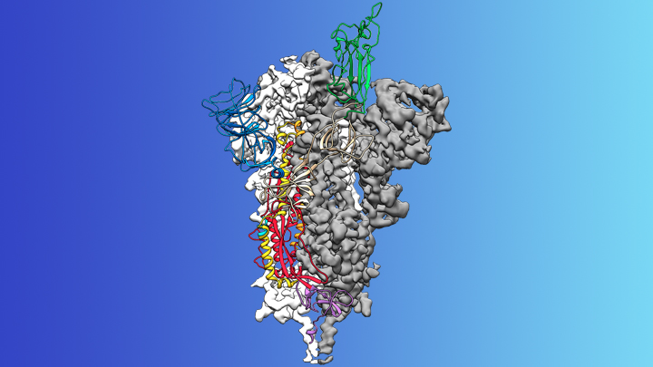
Credit: McLellan Lab, University of Texas at Austin
The recent COVID-19 outbreak of a novel type of coronavirus that began in China has prompted a massive global effort to contain and slow its spread. Despite those efforts, over the last month the virus has begun circulating outside of China in multiple countries and territories.
Cases have now appeared in the United States involving some affected individuals who haven’t traveled recently outside the country. They also have had no known contact with others who have recently arrived from China or other countries where the virus is spreading. The NIH and other U.S. public health agencies stand on high alert and have mobilized needed resources to help not only in its containment, but in the development of life-saving interventions.
On the treatment and prevention front, some encouraging news was recently reported. In record time, an NIH-funded team of researchers has created the first atomic-scale map of a promising protein target for vaccine development [1]. This is the so-called spike protein on the new coronavirus that causes COVID-19. As shown above, a portion of this spiky surface appendage (green) allows the virus to bind a receptor on human cells, causing other portions of the spike to fuse the viral and human cell membranes. This process is needed for the virus to gain entry into cells and infect them.
Preclinical studies in mice of a candidate vaccine based on this spike protein are already underway at NIH’s Vaccine Research Center (VRC), part of the National Institute of Allergy and Infectious Diseases (NIAID). An early-stage phase I clinical trial of this vaccine in people is expected to begin within weeks. But there will be many more steps after that to test safety and efficacy, and then to scale up to produce millions of doses. Even though this timetable will potentially break all previous speed records, a safe and effective vaccine will take at least another year to be ready for widespread deployment.
Coronaviruses are a large family of viruses, including some that cause “the common cold” in healthy humans. In fact, these viruses are found throughout the world and account for up to 30 percent of upper respiratory tract infections in adults.
This outbreak of COVID-19 marks the third time in recent years that a coronavirus has emerged to cause severe disease and death in some people. Earlier coronavirus outbreaks included SARS (severe acute respiratory syndrome), which emerged in late 2002 and disappeared two years later, and MERS (Middle East respiratory syndrome), which emerged in 2012 and continues to affect people in small numbers.
Soon after COVID-19 emerged, the new coronavirus, which is closely related to SARS, was recognized as its cause. NIH-funded researchers including Jason McLellan, an alumnus of the VRC and now at The University of Texas at Austin, were ready. They’d been studying coronaviruses in collaboration with NIAID investigators for years, with special attention to the spike proteins.
Just two weeks after Chinese scientists reported the first genome sequence of the virus [2], McLellan and his colleagues designed and produced samples of its spike protein. Importantly, his team had earlier developed a method to lock coronavirus spike proteins into a shape that makes them both easier to analyze structurally via the high-resolution imaging tool cryo-electron microscopy and to use in vaccine development efforts.
After locking the spike protein in the shape it takes before fusing with a human cell to infect it, the researchers reconstructed its atomic-scale 3D structural map in just 12 days. Their results, published in Science, confirm that the spike protein on the virus that causes COVID-19 is quite similar to that of its close relative, the SARS virus. It also appears to bind human cells more tightly than the SARS virus, which may help to explain why the new coronavirus appears to spread more easily from person to person, mainly by respiratory transmission.
McLellan’s team and his NIAID VRC counterparts also plan to use the stabilized spike protein as a probe to isolate naturally produced antibodies from people who’ve recovered from COVID-19. Such antibodies might form the basis of a treatment for people who’ve been exposed to the virus, such as health care workers.
The NIAID is now working with the biotechnology company Moderna, Cambridge, MA, to use the latest findings to develop a vaccine candidate using messenger RNA (mRNA), molecules that serve as templates for making proteins. The goal is to direct the body to produce a spike protein in such a way to elicit an immune response and the production of antibodies. An early clinical trial of the vaccine in people is expected to begin in the coming weeks. Other vaccine candidates are also in preclinical development.
Meanwhile, the first clinical trial in the U.S. to evaluate an experimental treatment for COVID-19 is already underway at the University of Nebraska Medical Center’s biocontainment unit [3]. The NIH-sponsored trial will evaluate the safety and efficacy of the experimental antiviral drug remdesivir in hospitalized adults diagnosed with COVID-19. The first participant is an American who was repatriated after being quarantined on the Diamond Princess cruise ship in Japan.
As noted, the risk of contracting COVID-19 in the United States is currently low, but the situation is changing rapidly. One of the features that makes the virus so challenging to stay in front of is its long latency period before the characteristic flu-like fever, cough, and shortness of breath manifest. In fact, people infected with the virus may not show any symptoms for up to two weeks, allowing them to pass it on to others in the meantime. You can track the reported cases in the United States on the Centers for Disease Control and Prevention’s website.
As the outbreak continues over the coming weeks and months, you can be certain that NIH and other U.S. public health organizations are working at full speed to understand this virus and to develop better diagnostics, treatments, and vaccines.
References:
[1] Cryo-EM structure of the 2019-nCoV spike in the prefusion conformation. Wrapp D, Wang N, Corbett KS, Goldsmith JA, Hsieh CL, Abiona O, Graham BS, McLellan JS. Science. 2020 Feb 19.
[2] A new coronavirus associated with human respiratory disease in China. Wu F, Zhao S, Yu B, Chen YM, Wang W, Song ZG, Hu Y, Tao ZW, Tian JH, Pei YY, Yuan ML, Zhang YL, Dai FH, Liu Y, Wang QM, Zheng JJ, Xu L, Holmes EC, Zhang YZ. Nature. 2020 Feb 3.
[3] NIH clinical trial of remdesivir to treat COVID-19 begins. NIH News Release. Feb 25, 2020.
Links:
Coronaviruses (National Institute of Allergy and Infectious Diseases/NIH)
Coronavirus (COVID-19) (NIAID)
Coronavirus Disease 2019 (Centers for Disease Control and Prevention, Atlanta)
NIH Support: National Institute of Allergy and Infectious Diseases