viral shedding
Seeing Coronavirus Replicate in Kidney Cells
Posted on by Dr. Francis Collins
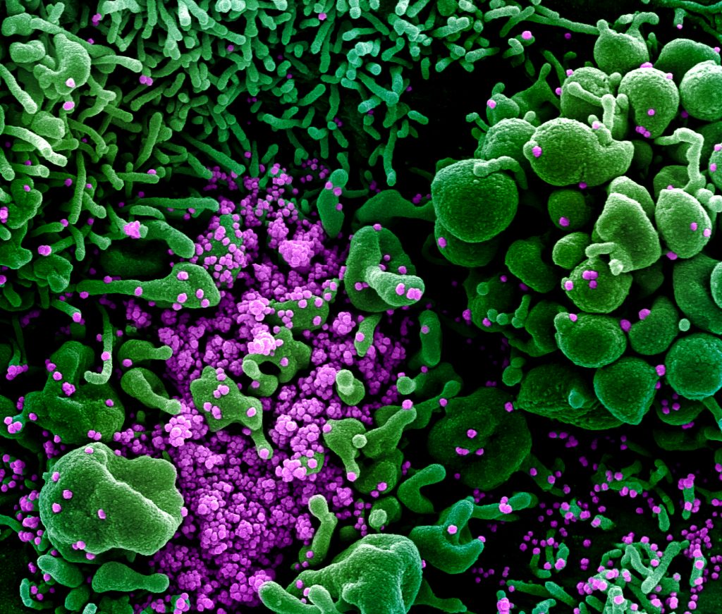
You’ve probably seen pictures of SARS-CoV-2—the novel coronavirus that causes COVID-19—that look alarming. But the high-resolution micrograph above paints a rather different picture, using rich pseudo-colors to show how newly assembled viral particles cause infected cells to bulge, or bleb, and then self-destruct.
This image depicts a common primate kidney cell line (green) infected with SARS-CoV-2. Notice the bulging, spherical cellular blebs, seen best in the upper right and bottom left corners. These badly damaged cells, which are filled to the point of bursting with viral particles, are beginning to self-destruct. Some cells have apparently already burst open, allowing hundreds of viral particles (purple) to spill out and potentially infect other cells.
This stunning picture was taken by John Bernbaum, an electron microscopist with NIH’s National Institute of Allergy and Infectious Diseases (NIAID). Bernbaum works at NIAID’s Integrated Research Facility (IRF), Fort Detrick, MD, a specialized, high-level biocontainment facility equipped with unique medical imaging capabilities. In this special environment, Bernbaum and his colleagues can safely visualize SARS-CoV-2, as well as other viruses and microbes that pose serious risks to human health.
To get this shot of SARS-CoV-2, Bernbaum relied on a conventional scanning electron microscope (SEM). First, a sample of kidney cells that had been exposed to SARS-CoV-2 was dehydrated, chemically preserved, and coated with a thin layer of metal. Once everything was ready, the SEM was used to focus a high-energy beam of electrons onto the sample. As electrons bounced off the metal surface, they revealed spatial variations and properties in the sample that were used to generate this 3D image.
Originally, this image was in gray scale. To better highlight the destructive powers of SARS-CoV-2, Jiro Wada, a skilled graphic illustrator at the IRF, used a computer program to colorize key features in exquisite detail. By studying these 3D images, researchers can learn about things such as the rate of infection and the prodigious number of particles each infected cell produces. They can also learn about how the infection affects the conditions inside cells.
Interestingly, what Bernbaum finds most striking about SARS-CoV-2 is what you don’t see in his images. Uninfected kidney cells look like a flat, delicately interwoven quilt (not pictured). When Bernbaum used SEM to study this sample of kidney cells, about 80 to 90 percent of the cells appeared flat and unremarkable. Yet, as the scan progressed, he came across a small subset of cells that appeared to be deformed by SARS-CoV-2 infection. Those abnormalities include the spherical bulges that I pointed out earlier, along with some worm-like protrusions that you can see in the top left.
Bernbaum has been producing amazing images like this one for 32 years—the last 11 of them at the IRF. If you’d like to see even more of his impressive work and that of the IRF team, check out the NIAID’s image gallery.
Links:
Coronavirus (NIH)
Integrated Research Facility (National Institute of Allergy and Infectious Diseases/NIH)
NIH Support: National Institute of Allergy and Infectious Diseases
Capturing Viral Shedding in Action
Posted on by Dr. Francis Collins

You’ve probably seen some amazing high-resolution images of SARS-CoV-2, the novel coronavirus that causes COVID-19, on television and the web. What you might not know is that many of these images, including the ones shown here, were produced at Rocky Mountain Laboratories (RML), a part of NIH’s National Institute of Allergy and Infectious Diseases (NIAID) that’s located in the small Montana town of Hamilton.
The head of RML’s Electron Microscopy Unit, Elizabeth Fischer, was the researcher who took this portrait of SARS-CoV-2. For more than 25 years, Fischer has snapped stunning images of dangerous viruses and microbes, including some remarkable shots of the deadly Ebola virus. She also took some of the first pictures of the coronavirus that causes Middle East respiratory syndrome (MERS), which arose from camels and continues to circulate at low levels in people.
The NIAID facility uses a variety of microscopy techniques, including state-of-the-art cryo-electron microscopy (cryo-EM). But the eye-catching image you see here was taken with a classic scanning electron microscope (SEM).
SEM enables visualization of particles, including viruses, that are too small to be seen with traditional light microscopy. It does so by focusing electrons, instead of light, into a beam that scans the surface of a sample that’s first been dehydrated, chemically preserved, and then coated with a thin layer of metal. As electrons bounce off the sample’s surface, microscopists such as Fischer are able to capture its precise topology. The result is a gray-scale micrograph like the one you see above on the left. To make the image easier to interpret, Fischer hands the originals off to RML’s Visual Medical Arts Department, which uses colorization to make key features pop like they do in the image on the right.
So, what exactly are you seeing in this image? The orange-brown folds and protrusions are part of the surface of a single cell that’s been infected with SARS-CoV-2. This particular cell comes from a commonly studied primate kidney epithelial cell line. The small, blue spheres emerging from the cell surface are SARS-CoV-2 particles.
This picture is quite literally a snapshot of viral shedding, a process in which viral particles are released from a dying cell. This image gives us a window into how devastatingly effective SARS-CoV-2 appears to be at co-opting a host’s cellular machinery: just one infected cell is capable of releasing thousands of new virus particles that can, in turn, be transmitted to others.
While capturing a fixed sample on the microscope is fairly straightforward for a pro like Fischer, developing a sample like this one involves plenty of behind-the-scenes trial and error by NIAID investigators. As you might imagine, to see the moment that viruses emerge from an infected cell, you have to get the timing just right.
By capturing many shots of the coronavirus using the arsenal of microscopes available at RML and elsewhere, researchers are learning more every day about how SARS-CoV-2 enters a cell, moves inside it, and then emerges to infect other cells. In addition to advancing scientific knowledge, Fischer notes that images like these also hold the remarkable power to make an invisible enemy visible to the world at large.
Making SARS-CoV-2 tangible helps to demystify the challenges that all of us now face as a result of the COVID-19 pandemic. The hope is it will encourage each and every one of us to do our part to fight it, whether that means digging into the research, working on the front lines, or staying at home to prevent transmission and flatten the curve. And, if you could use some additional inspiration, don’t miss the NIAID’s image gallery on Flickr, which includes some of Fischer’s finest work.
Links:
Coronavirus (COVID-19) (NIH)
Rocky Mountain Laboratories (National Institute of Allergy and Infectious Diseases/NIH)
Elizabeth Fischer (National Institute of Allergy and Infectious Diseases/NIH)
NIH Support: National Institute of Allergy and Infectious Diseases