blebbing
Seeing Coronavirus Replicate in Kidney Cells
Posted on by Dr. Francis Collins
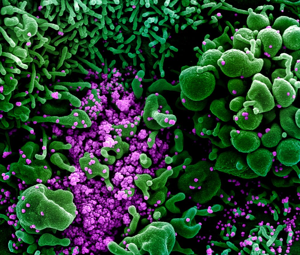
You’ve probably seen pictures of SARS-CoV-2—the novel coronavirus that causes COVID-19—that look alarming. But the high-resolution micrograph above paints a rather different picture, using rich pseudo-colors to show how newly assembled viral particles cause infected cells to bulge, or bleb, and then self-destruct.
This image depicts a common primate kidney cell line (green) infected with SARS-CoV-2. Notice the bulging, spherical cellular blebs, seen best in the upper right and bottom left corners. These badly damaged cells, which are filled to the point of bursting with viral particles, are beginning to self-destruct. Some cells have apparently already burst open, allowing hundreds of viral particles (purple) to spill out and potentially infect other cells.
This stunning picture was taken by John Bernbaum, an electron microscopist with NIH’s National Institute of Allergy and Infectious Diseases (NIAID). Bernbaum works at NIAID’s Integrated Research Facility (IRF), Fort Detrick, MD, a specialized, high-level biocontainment facility equipped with unique medical imaging capabilities. In this special environment, Bernbaum and his colleagues can safely visualize SARS-CoV-2, as well as other viruses and microbes that pose serious risks to human health.
To get this shot of SARS-CoV-2, Bernbaum relied on a conventional scanning electron microscope (SEM). First, a sample of kidney cells that had been exposed to SARS-CoV-2 was dehydrated, chemically preserved, and coated with a thin layer of metal. Once everything was ready, the SEM was used to focus a high-energy beam of electrons onto the sample. As electrons bounced off the metal surface, they revealed spatial variations and properties in the sample that were used to generate this 3D image.
Originally, this image was in gray scale. To better highlight the destructive powers of SARS-CoV-2, Jiro Wada, a skilled graphic illustrator at the IRF, used a computer program to colorize key features in exquisite detail. By studying these 3D images, researchers can learn about things such as the rate of infection and the prodigious number of particles each infected cell produces. They can also learn about how the infection affects the conditions inside cells.
Interestingly, what Bernbaum finds most striking about SARS-CoV-2 is what you don’t see in his images. Uninfected kidney cells look like a flat, delicately interwoven quilt (not pictured). When Bernbaum used SEM to study this sample of kidney cells, about 80 to 90 percent of the cells appeared flat and unremarkable. Yet, as the scan progressed, he came across a small subset of cells that appeared to be deformed by SARS-CoV-2 infection. Those abnormalities include the spherical bulges that I pointed out earlier, along with some worm-like protrusions that you can see in the top left.
Bernbaum has been producing amazing images like this one for 32 years—the last 11 of them at the IRF. If you’d like to see even more of his impressive work and that of the IRF team, check out the NIAID’s image gallery.
Links:
Coronavirus (NIH)
Integrated Research Facility (National Institute of Allergy and Infectious Diseases/NIH)
NIH Support: National Institute of Allergy and Infectious Diseases
Snapshots of Life: Biological Bubble Machine
Posted on by Dr. Francis Collins

Credit: Chi Zhao, David Busch, Connor Vershel, Jeanne Stachowiak, University of Texas at Austin
As kids, most of us got a bang out of blowing soap bubbles and watching them float around. Biologists have learned that some of our cells do that too. On the right, you can see two cells (greenish yellow) in the process of forming bubbles, or plasma membrane vesicles (PMVs). During this blebbing process, a cell’s membrane temporarily disassociates from its underlying cytoskeleton, forming a tiny pouch that, over the course of about 30 minutes, is “inflated” with a mix of proteins and lipids from inside the cell. After the PMVs are fully filled, these bubble-like structures are pinched off and released, like those that you see in the background. Certain cells constantly release PMVs, along with other types of vesicles, and may use those to communicate with other cells throughout the body.
This particular image, an entrant in the Biophysical Society’s 2017 Art of Science Image Contest, was produced by researchers working in the NIH-supported lab of Jeanne Stachowiak at the University of Texas at Austin. Stachowiak’s group is among the first to explore the potential of PMVs as specialized drug-delivery systems to target cancer and other disorders [1].
Until recently, most efforts to exploit vesicles for therapeutic uses have employed synthetic versions of a different type of vesicle, called an exosome. But Stachowiak and others have realized that PMVs come with certain built-in advantages. A major one is that a patient’s own cells could in theory serve as the production facility.