FASEB Bioart 2017
Using Frogs to Tackle Kidney Problems
Posted on by Dr. Francis Collins
Many human cells are adorned with hair-like projections called cilia. Scientists now realize that these specialized structures play many important roles throughout the body, including directing or sensing various signals such as fluid flow. Their improper function has been linked to a wide range of health conditions, such as kidney disease, scoliosis, and obesity.
Studying cilia in people can be pretty challenging. It’s less tricky in a commonly used model organism: Xenopus laevis, or the African clawed frog. This image highlights a healthy patch of motile cilia (yellow) on embryonic skin cells (red) of Xenopus laevis. The cilia found in humans and all other vertebrates are built from essentially the same elongated structures known as microtubules. That’s why researchers can learn a lot about human cilia by studying frogs.
A Microbial Work of Art
Posted on by Dr. Francis Collins
Credit: Scott Chimileski, Sylvie Laborde, Nicholas Lyons, Roberto Kolter, Harvard Medical School, Boston
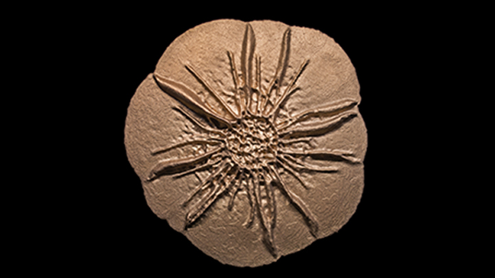
Bacteria are single-celled organisms that are too small to see in detail without the aid of a microscope. So you might not think that zooming in on a batch of bacteria would provide the inspiration for a museum-worthy sculpture.
But, in fact, that’s exactly what you see in the image. Researchers grew in a lab dish Bacillus licheniformis, a usually benign bacterium from the soil that produces an enzyme used in laundry detergent. The bacteria self-organized into a sand dollar-like pattern to form a cohesive structure called a biofilm. The researchers then took a 3D scan of the living bacterial colony in the lab and used it to print this stainless steel sculpture at 12 times the dime-sized biofilm.
First Day in the Life of Nine Amazing Creatures
Posted on by Dr. Francis Collins
Credit: Tessa Montague, Harvard University, and Zuzka Vavrušová, University of California, San Francisco
Each summer for the last 125 years, students from around the country have traveled to the Marine Biological Laboratory (MBL), Woods Hole, MA, for an intensive course in embryology. While visiting this peaceful and scenic village on Cape Cod, they’re exposed to a dizzying array of organisms and state-of-the-art techniques to study their development.
A Lean, Mean DNA Packaging Machine
Posted on by Dr. Francis Collins
Credit: Victor Padilla-Sanchez, The Catholic University of America, Washington, D.C.
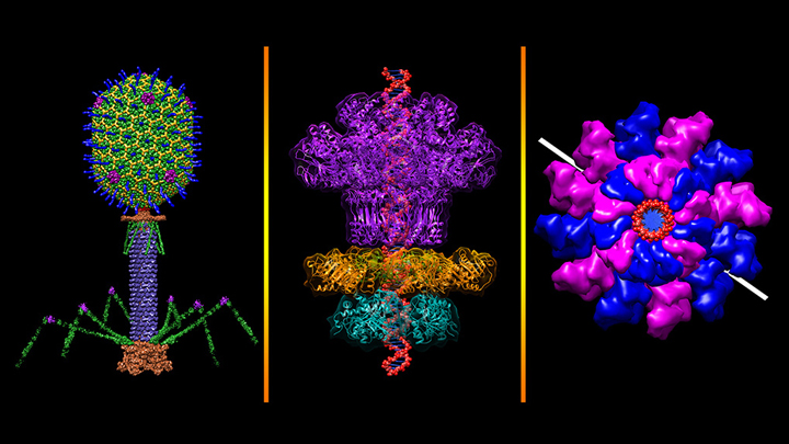
All plants and animals are susceptible to viral infections. But did you know that’s also true for bacteria? They get nailed by viruses called bacteriophages, and there are thousands of them in nature including this one that resembles a lunar lander: bacteriophage T4 (left panel). It’s a popular model organism that researchers have studied for nearly a century, helping them over the years to learn more about biochemistry, genetics, and molecular biology [1].
The bacteriophage T4 infects the bacterium Escherichia coli, which normally inhabits the gastrointestinal tract of humans. T4’s invasion starts by touching down on the bacterial cell wall and injecting viral DNA through its tube-like tail (purple) into the cell. A DNA “packaging machine” (middle and right panels) between the bacteriophage’s “head” and “tail” (green, yellow, blue spikes) keeps the double-stranded DNA (middle panel, red) at the ready. All the vivid colors you see in the images help to distinguish between the various proteins or protein subunits that make up the intricate structure of the bacteriophage and its DNA packaging machine.
Red Blood Cells and Mercury
Posted on by Dr. Francis Collins
Credit: Courtney Fleming, Birnur Akkaya, and Umut Gurkan, Case Western Reserve University, Cleveland
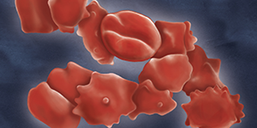
Mercury is a naturally occurring heavy metal and a well-recognized environmental toxin. When absorbed into the bloodstream at elevated levels, mercury is also extremely harmful to people, causing a range of problems including cognitive impairments, skin rashes, and kidney problems [1].
In this illustration, it’s possible to see in red blood cells the effects of mercury chloride, a toxic chemical compound now sometimes used as a laboratory reagent. Normally, healthy red blood cells have a distinct, doughnut-like shape that helps them squeeze through the tiniest of blood vessels. But these cells are terribly disfigured, with unusual spiky projections, after 24 hours of exposure to low levels of a mercury chloride in solution.
Snapshots of Life: Building Muscle in a Dish
Posted on by Dr. Francis Collins

Credit: Kevin Murach, Charlotte Peterson, and John McCarthy, University of Kentucky, Lexington
As many of us know from hard experience, tearing a muscle while exercising can be a real pain. The good news is that injured muscle will usually heal quickly for many of us with the help of satellite cells. Never heard of them? They are the adult stem cells in our skeletal muscles long recognized for their capacity to make new muscle fibers called myotubes.
This striking image shows what happens when satellite cells from mice are cultured in a lab dish. With small adjustments to the lab dish’s growth media, those cells fuse to form myotubes. Here, you see the striated myotubes (red) with multiple cell nuclei (blue) characteristic of mature muscle fibers. The researchers also used a virus to genetically engineer some of the muscle to express a fluorescent protein (green).
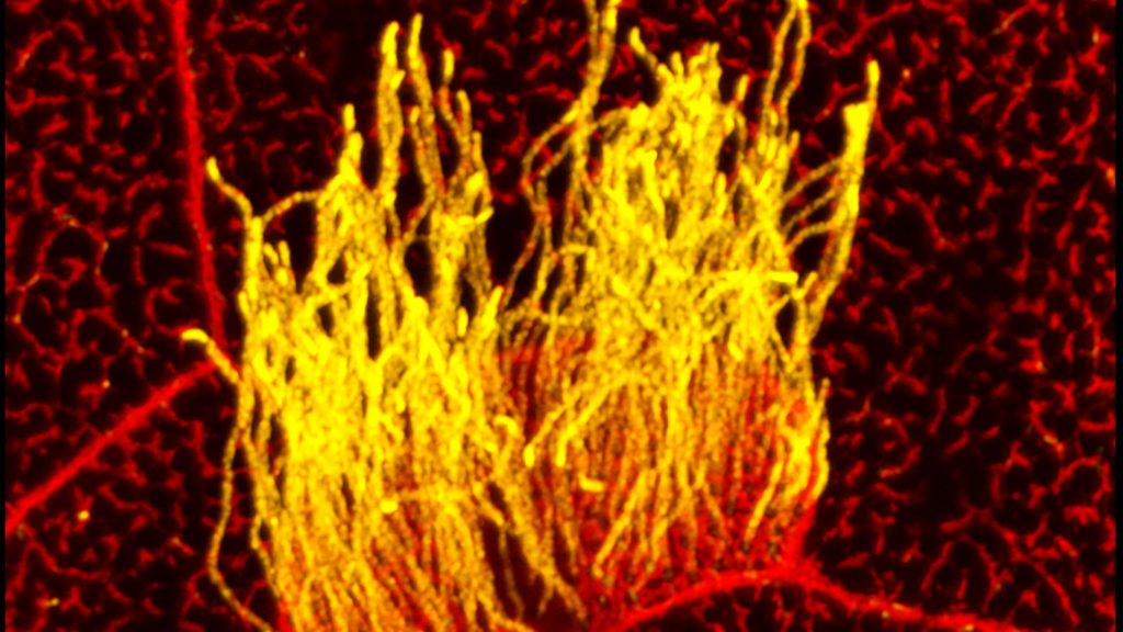
Snapshots of Life: The Brain’s Microscopic Green Trash Bins
Posted on by Dr. Francis Collins
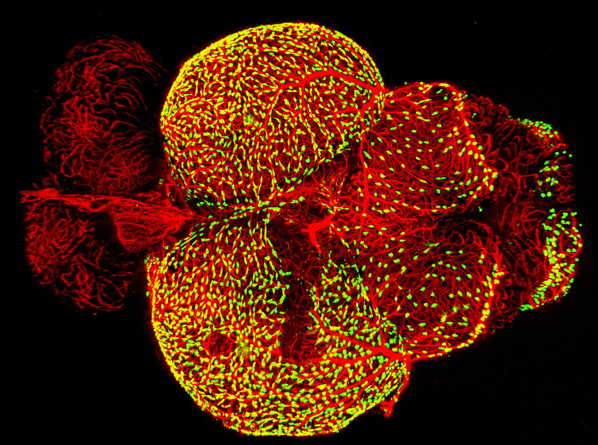
Credit: Marina Venero Galanternik, Daniel Castranova, Tuyet Nguyen, and Brant M. Weinstein, NICHD, NIH
There are trash bins in our homes, on our streets, and even as a popular icon on our desktop computers. And as this colorful image shows, trash bins of the cellular variety are also important in the brain.
This image—a winner in the Federation of American Societies for Experimental Biology’s 2017 BioArt competition—shows the brain of an adult zebrafish, a popular organism for studying how the brain works. It captures dense networks of blood vessels (red) lining the outer surface of the brain. Next to many of these vessels sit previously little-studied cells called fluorescent granular perithelial cells (yellowish green). Researchers now believe these cells, often shortened to FGPs, act much like trash receptacles that continuously take in and store waste products to keep the brain tidy and functioning well.