fluorescent granular perithelial cells
Snapshots of Life: The Brain’s Microscopic Green Trash Bins
Posted on by Dr. Francis Collins
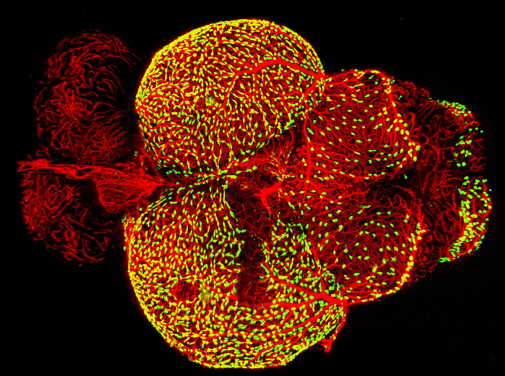
Credit: Marina Venero Galanternik, Daniel Castranova, Tuyet Nguyen, and Brant M. Weinstein, NICHD, NIH
There are trash bins in our homes, on our streets, and even as a popular icon on our desktop computers. And as this colorful image shows, trash bins of the cellular variety are also important in the brain.
This image—a winner in the Federation of American Societies for Experimental Biology’s 2017 BioArt competition—shows the brain of an adult zebrafish, a popular organism for studying how the brain works. It captures dense networks of blood vessels (red) lining the outer surface of the brain. Next to many of these vessels sit previously little-studied cells called fluorescent granular perithelial cells (yellowish green). Researchers now believe these cells, often shortened to FGPs, act much like trash receptacles that continuously take in and store waste products to keep the brain tidy and functioning well.