cleft palate
Halloween Fly-Through of a Mouse Skull
Posted on by Dr. Francis Collins
Credit: Chai Lab, University of Southern California, Los Angeles
Halloween is full of all kinds of “skulls”—from spooky costumes to ghoulish goodies. So, in keeping with the spirit of the season, I’d like to share this eerily informative video that takes you deep inside the real thing.
Snapshots of Life: Coming Face to Face with Development
Posted on by Dr. Francis Collins
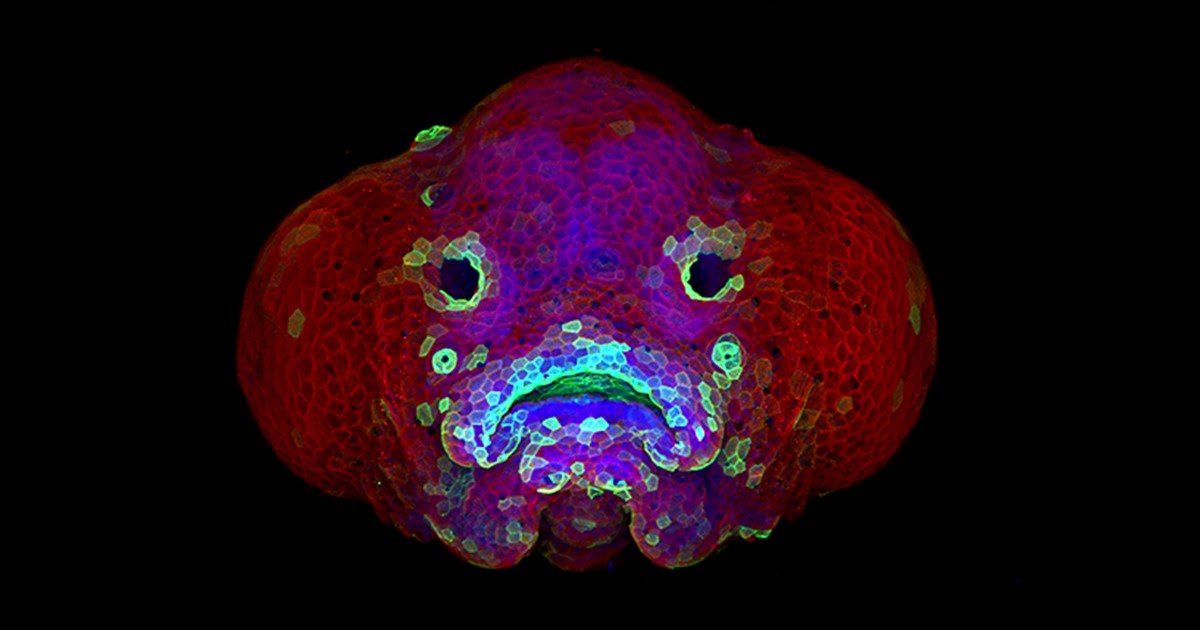
Credit: Oscar Ruiz and George Eisenhoffer, University of Texas MD Anderson Cancer Center, Houston
Zebrafish (Danio rerio) is a favorite model for studying development, in part because its transparent embryos make it possible to produce an ever-growing array of amazingly informative images. For one recent example, check out this Federation of American Societies for Experimental Biology’s 2016 BioArt winner, which shows the developing face of a 6-day-old zebrafish larva.
Yes, those downturned “lips” are indeed cells that will go on to become the fish’s mouth. But all is not quite what it appears: the two dark circles that look like eyes are actually developing nostrils. Both the nostrils and mouth express high levels of F-actin (green), a structural protein that helps orchestrate cell movement. Meanwhile, the two bulging areas on either side of the fish’s head, which are destined to become eyes and skin, express keratin (red).
Oscar Ruiz, who works in the lab of George Eisenhoffer at The University of Texas MD Anderson Cancer Center, Houston, used a confocal microscope to create this image. What was most innovative about his work was not the microscope itself, but how he prepared the sample for imaging. With traditional methods, researchers can only image the faces of zebrafish larvae from the side or the bottom. However, the Eisenhoffer lab has devised a new method of preparing fish larvae that makes it possible to image their faces head-on. This has enabled the team to visualize facial development at much higher resolution than was previously possible.
Snapshots of Life: Development in Exquisite Detail
Posted on by Dr. Francis Collins
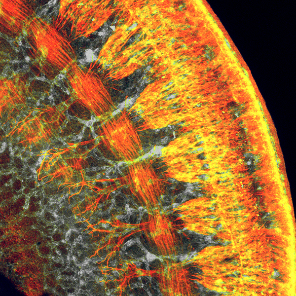
Credit: Shachi Bhatt and Paul Trainor, Stowers Institute for Medical Research, Kansas City, MO
If you’ve ever tried to take photos of wiggly kids, you know that it usually takes several attempts before you get the perfect shot. It’s often the same for biomedical researchers when taking images with microscopes because there are so many variables—from sample preparation to instrument calibration—to take into account. Still, there are always exceptions where everything comes together just right, and you are looking at one of them! On her first try at using a confocal microscope to image this cross-section of a mouse embryo’s torso, postdoc Shachi Bhatt captured a gem of an image that sheds new light on mammalian development.
Bhatt, who works in the NIH-supported lab of Paul Trainor at the Stowers Institute for Medical Research, Kansas City, MO, produced this micrograph as part of a quest to understand the striking parallels seen between the development of the nervous system and the vascular system in mammals. Fluorescent markers were used to label proteins uniquely expressed in each type of tissue: reddish-orange delineates developing nerve cells; gray highlights developing blood vessels; and yellow shows where the nerve cells and blood vessels overlap.
Largest Study Yet Shows Mother’s Smoking Changes Baby’s Epigenome
Posted on by Dr. Francis Collins

Credit: Daniel Berehulak/Getty Images
Despite years of public health campaigns warning of the dangers of smoking when pregnant, many women are unaware of the risk or find themselves unable to quit. As a result, far too many babies are still being exposed in the womb to toxins that enter their mothers’ bloodstreams when they inhale cigarette smoke. Among the many infant and child health problems that have been linked to maternal smoking are premature birth, low birth weight, asthma, reduced lung function, sudden infant death syndrome (SIDS), and cleft lip and/or palate.
Now, a large international study involving NIH-supported researchers provides a biological mechanism that may explain how exposure to cigarette toxins during fetal development can produce these health problems [1]. That evidence centers on the impact of the toxins on the epigenome of the infant’s body tissues. The epigenome refers to chemical modifications of DNA (particularly methylation of cytosines), as well as proteins that bind to DNA and affect its function. The genome of an individual is the same in all cells of their body, but the epigenome determines whether genes are turned on or off in particular cells. The study found significant differences between the epigenetic patterns of babies born to women who smoked during pregnancy and those born to non-smokers, with many of the differences affecting genes known to play key roles in the development of the lungs, face, and nervous system.