epithelium
Possible Explanation for Why Some People Get More Colds
Posted on by Dr. Francis Collins

Getty Images/yourstockbank
Colds are just an occasional nuisance for many folks, but some individuals seem to come down with them much more frequently. Now, NIH-funded researchers have uncovered some new clues as to why.
In their study, the researchers found that the cells that line our airways are quite adept at defending against cold-causing rhinoviruses. But there’s a tradeoff. When these cells are busy defending against tissue damage due to cigarette smoke, pollen, pollutants, and/or other airborne irritants, their ability to fend off such viruses is significantly reduced [1].
The new findings may open the door to better strategies for preventing the common cold, as well as other types of respiratory tract infections caused by non-flu viruses. Even small improvements in prevention could have big implications for our nation’s health and economy. Every year, Americans come down with more than 500 million colds and similar infections, leading to reduced work productivity, medical expenses, and other costs approaching $40 billion [2].
Snapshots of Life: Coming Face to Face with Development
Posted on by Dr. Francis Collins
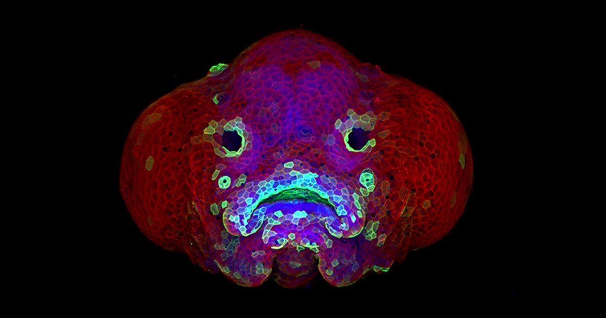
Credit: Oscar Ruiz and George Eisenhoffer, University of Texas MD Anderson Cancer Center, Houston
Zebrafish (Danio rerio) is a favorite model for studying development, in part because its transparent embryos make it possible to produce an ever-growing array of amazingly informative images. For one recent example, check out this Federation of American Societies for Experimental Biology’s 2016 BioArt winner, which shows the developing face of a 6-day-old zebrafish larva.
Yes, those downturned “lips” are indeed cells that will go on to become the fish’s mouth. But all is not quite what it appears: the two dark circles that look like eyes are actually developing nostrils. Both the nostrils and mouth express high levels of F-actin (green), a structural protein that helps orchestrate cell movement. Meanwhile, the two bulging areas on either side of the fish’s head, which are destined to become eyes and skin, express keratin (red).
Oscar Ruiz, who works in the lab of George Eisenhoffer at The University of Texas MD Anderson Cancer Center, Houston, used a confocal microscope to create this image. What was most innovative about his work was not the microscope itself, but how he prepared the sample for imaging. With traditional methods, researchers can only image the faces of zebrafish larvae from the side or the bottom. However, the Eisenhoffer lab has devised a new method of preparing fish larvae that makes it possible to image their faces head-on. This has enabled the team to visualize facial development at much higher resolution than was previously possible.