Life: Magnified
Snapshots of Life: Cell Skeleton on the Move
Posted on by Dr. Francis Collins

Credit: Torsten Wittmann, University of California, San Francisco
Cells are constantly on the move. They shift, grow, and migrate to new locations—for example, to heal a wound or to intercept an infectious agent as part of an immune response. But how do cells actually move?
In this image, Torsten Wittmann, an NIH-funded cell biologist at the University of California, San Francisco, reveals the usually-invisible cytoskeleton of a normal human skin cell that lends the cell its mobility. The cytoskeleton is made from protein structures called microtubules—the wispy threads surrounding the purple DNA-containing nucleus—and filaments of a protein called actin, seen here as the fine blue meshwork in the cell periphery. Both actin and microtubules are critical for growth and movement.
Snapshots of Life: Visualizing Blood Vessels
Posted on by Dr. Francis Collins

Credit: Christopher V. Carman and Roberta Martinelli, Harvard Medical School, Boston
This might look a bit like a fish net, but what’s actually caught in this image is the structure of the endothelium—the thin layer of cells lining your blood vessels that controls the flow of molecules in and out of the bloodstream. The red lines are the actin filaments that give each endothelial cell its shape, while the purple are proteins called cadherins.
Most of the time, the actin “ropes” and cadherin “glue” act together to form a tight seal between endothelial cells, ensuring that nothing leaks out of blood vessels into surrounding tissue. However, when endothelial cells sense an infection or an injury, the cadherins open gaps that allow various disease-fighting or healing factors or cells present in the blood to breach the barrier and enter infected or injured tissue. After the infection subsides or wound heals, the gaps close and the blood vessel is once again impenetrable.
Snapshots of Life: Fat (Tissue) Is Beautiful
Posted on by Dr. Francis Collins
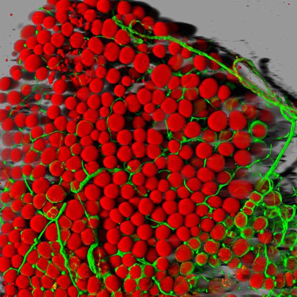
Caption: Fat cells (red) surrounded by blood vessels (green) that supply them with nutrients.
Credit: Daniela Malide, National Heart, Lung, and Blood Institute; NIH
With all of today’s sophisticated microscopes, you’d think it would be simple to take high-magnification photos of fat—but it’s not. Fat tissue often leaks slippery contents, namely lipids, when it’s thinly sliced for viewing under a microscope. And even when a sample is prepared without leakage, there’s another hurdle: the viscous droplets of lipid contained in the fat cells block light from passing through.
So, it’s good news that one of NIH’s intramural scientists here in Bethesda, MD, has come up with a way to produce high-resolution, 3-D images of fat cells like the one you see above. Not only are these images aesthetically appealing, but they’ll be valuable to efforts to expand our understanding of this essential and much-maligned tissue.
Snapshots of Life: Behold the Beauty of the Eye
Posted on by Dr. Francis Collins

Credit: Bryan William Jones and Robert E. Marc, University of Utah
The eye is a complex marvel of nature. In fact, there are some 70 to 80 kinds of cells in the mammalian retina. This image beautifully illuminates the eye’s complexity, on a cellular level—showing how these cells are arranged and wired together to facilitate sight.
“Reading” the image from left to right, we first find the muscle cells, in peach, that move the eye in its socket. The green layer, next, is the sclera—the white part of the eye. The spongy-looking layers that follow provide blood to the retina. The thin layer of yellow is the retinal pigment epithelium. The photoreceptors, in shades of pink, detect photons and transmit the information to the next layer down: the bipolar and horizontal cells (purple). From the bipolar cells, information flows to the amacrine and ganglion cells (blue, green, and turquoise) and then out of the retina via the optic nerve (the white plume that seems to billow out across the upper-right side of the eye), which transmits data to the brain for processing.
Snapshots of Life: Wild Outcome from Knocking Out Mobility Proteins
Posted on by Dr. Francis Collins

Credit: Praveen Suraneni and Rong Li, Stowers Institute for Medical Research
When biologists disabled proteins critical for cell movement, the result was dramatic. The membrane, normally a smooth surface enveloping the cell, erupted in spiky projections. This image, which is part of the Life: Magnified exhibit, resembles a supernova. Although it looks like it exploded, the cell pictured is still alive.
To create the image, Rong Li and Praveen Suraneni, NIH-funded cell biologists at the Stowers Institute for Medical Research in Kansas City, Missouri, disrupted two proteins essential to movement in fibroblasts—connective tissue cells that are also important for healing wounds. The first, called ARPC3, is a protein in the Arp2/3 complex. Without it, the cell moves more slowly and randomly [1]. Inhibiting the second protein gave this cell its spiky appearance. Called myosin IIA (green in the image), it’s like the cell’s muscle, and it’s critical for movement. The blue color is DNA; the red represents a protein called F-actin.
Snapshots of Life: Portrait of Skin Cancer
Posted on by Dr. Francis Collins
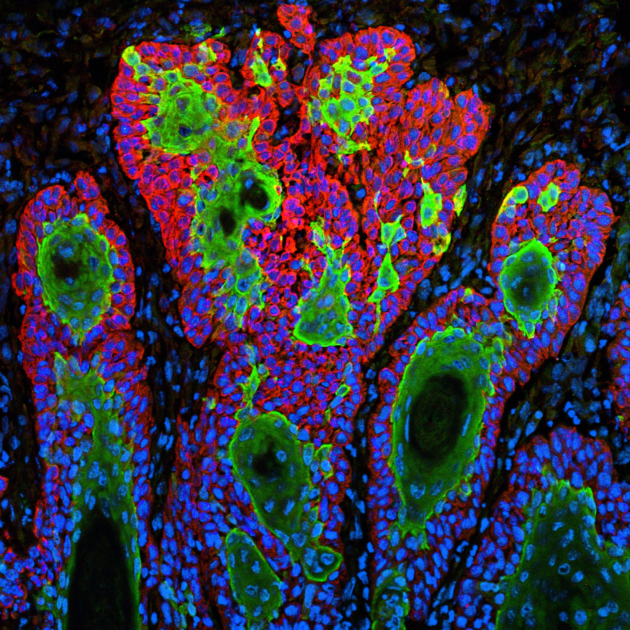
Caption: This image shows the uncontrolled growth of cells in squamous cell carcinoma.
Credit: Markus Schober and Elaine Fuchs, The Rockefeller University, New York
For Markus Schober, science is more inspiring when the images are beautiful, even when the subject is not. So, when this biologist was at The Rockefeller University in New York and peered through his microscope at squamous cell carcinoma (SCC), both the diabolical complexity—and the beauty—of this common form of skin cancer caught his eye.
Schober wasn’t the only one who found the image compelling. A panel of judges from the National Institute of General Medical Sciences and the American Society for Cell Biology chose to feature it in their Life: Magnified exhibit, which recently opened at the Washington Dulles International Airport.