LipidTox Red
Snapshots of Life: Fat (Tissue) Is Beautiful
Posted on by Dr. Francis Collins
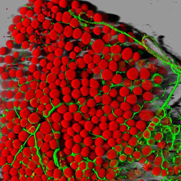
Caption: Fat cells (red) surrounded by blood vessels (green) that supply them with nutrients.
Credit: Daniela Malide, National Heart, Lung, and Blood Institute; NIH
With all of today’s sophisticated microscopes, you’d think it would be simple to take high-magnification photos of fat—but it’s not. Fat tissue often leaks slippery contents, namely lipids, when it’s thinly sliced for viewing under a microscope. And even when a sample is prepared without leakage, there’s another hurdle: the viscous droplets of lipid contained in the fat cells block light from passing through.
So, it’s good news that one of NIH’s intramural scientists here in Bethesda, MD, has come up with a way to produce high-resolution, 3-D images of fat cells like the one you see above. Not only are these images aesthetically appealing, but they’ll be valuable to efforts to expand our understanding of this essential and much-maligned tissue.