cancer cells
Cool Videos: Spying on Cancer Cell Invasion
Posted on by Dr. Francis Collins

If you’re a fan of the Mission: Impossible spy thrillers, you might think that secret agent Ethan Hunt has done it all. But here’s a potentially life-saving mission that his force has yet to undertake: spying on cancer cells. Never fear—some scientific sleuths already have!
So, have a look at this bio-action flick recently featured in the American Society for Cell Biology’s 2015 Celldance video series. Without giving too much of the plot away, let me just say that it involves cancer cells escaping from a breast tumor and spreading, or metastasizing, to other parts of the body. Along the way, those dastardly cancer cells take advantage of collagen fibers to make a tight-rope getaway and recruit key immune cells, called macrophages, to serve as double agents to aid and abet their diabolical spread.
Cool Videos: Cytotoxic T Cells on Patrol
Posted on by Dr. Francis Collins
Wow! It’s one thing to know that the immune system has the power to destroy cancerous cells. But it’s quite another thing to see a cytotoxic T cell actually take out a cancer cell right before your eyes.
This amazing video was produced by Alex T. Ritter as part of Celldance 2014, an annual video series by the American Society for Cell Biology (ASCB). To make this series happen in 2014, ASCB staff contacted cell biology labs known for their sophisticated imaging tools and techniques, asking them to submit proposals for videos. In return, ASCB provided some funding, post-production support from a professional videographer, and an original soundtrack from the up-and-coming Hollywood composer Ted Masur.
Snapshots of Life: Wild Outcome from Knocking Out Mobility Proteins
Posted on by Dr. Francis Collins

Credit: Praveen Suraneni and Rong Li, Stowers Institute for Medical Research
When biologists disabled proteins critical for cell movement, the result was dramatic. The membrane, normally a smooth surface enveloping the cell, erupted in spiky projections. This image, which is part of the Life: Magnified exhibit, resembles a supernova. Although it looks like it exploded, the cell pictured is still alive.
To create the image, Rong Li and Praveen Suraneni, NIH-funded cell biologists at the Stowers Institute for Medical Research in Kansas City, Missouri, disrupted two proteins essential to movement in fibroblasts—connective tissue cells that are also important for healing wounds. The first, called ARPC3, is a protein in the Arp2/3 complex. Without it, the cell moves more slowly and randomly [1]. Inhibiting the second protein gave this cell its spiky appearance. Called myosin IIA (green in the image), it’s like the cell’s muscle, and it’s critical for movement. The blue color is DNA; the red represents a protein called F-actin.
DNA Barcodes Interrogate Cancer Cells
Posted on by Dr. Francis Collins

Caption: A mix of cells collected from an abdominal cancer. The cancer cells (green) are positive for a cell surface cancer marker called EpCAM. The red cell is a normal mesothelial cell. The nuclei of all the cells are stained blue. Each of the five rows in the red, orange, and yellow “heat map” in the corner represents one cell, and the intensity of the color in each of the ~30 narrow columns reflects the abundance of a particular protein. It is apparent that there is a lot of heterogeneity in this collection of cancer cells.
Credit: Ralph Weissleder, Center for Systems Biology, Massachusetts General Hospital, Boston
The proteins speckling the surface of a cancer cell reveal critical clues—the type of cancer cell and a menu of possible mutations that may have triggered the malignancy. Since these proteins are exposed on the outside of the cell, they are also ideal targets for so-called precision cancer therapies (especially monoclonal antibodies), optimized for the particular individual. But in the past, to analyze and identify these different proteins, large samples of tissue have been needed. Typically, these are derived from surgical biopsies. But biopsies are expensive and invasive. Furthermore, they aren’t a practical option if you want to monitor the effects of a drug in a patient closely over time.
Using a minimally invasive method of cell sampling called fine needle aspiration, physicians can collect miniscule cell samples frequently, cheaply, and safely. But, until now, these tiny samples only provided enough material to analyze a handful of cell surface proteins. So, it comes as particularly good news that NIH-funded researchers at Massachusetts General Hospital in Boston have developed a new technology that quickly identifies hundreds of these proteins simultaneously, using just a few of the patient’s cells [1]. The key to this new method is a clever adaptation of the familiar barcode.
Human Folate Receptor Model May Aid Antifolate Drug Design
Posted on by Dr. Francis Collins
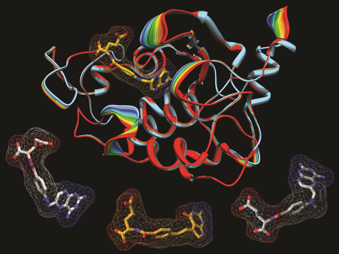
Caption: A model of the human folate receptor (top) and three antifolate drugs used in chemotherapy: aminopterin (left), pemetrexed, and methotrexate (right).
Credit: Charles Dann III / Courtesy of Indiana University
Vitamin B9 or folic acid, which is found in dark green leafy vegetables, is essential for cells to grow and divide rapidly—as they do in a growing embryo. This is why women are advised to take folic acid supplements before conception and during pregnancy: inadequate folate raises the risk of brain and spinal cord defects. But while folic acid is key to normal cell growth, rapidly dividing cancer cells also have a tremendous appetite for this vitamin.
Drugs called antifolates have been used for decades in chemotherapy to starve cancer cells of folate, which can help kill the tumor. These drugs have also been used to treat inflammatory diseases like rheumatoid arthritis and Crohn’s disease. But many of these drugs have nasty side effects because they also enter normal healthy cells, depriving them of this essential compound.
exRNA: Helping Cells Get Their Message Out
Posted on by Dr. Francis Collins

Source: NIH Common Fund
When your email is interrupted or blocked, it creates havoc. Messages remain undelivered, stalling interactions between you and your friends, family, and colleagues at work. Likewise when communication fails between your body’s cells, disease can result. Scientists recently discovered a new group of molecules called extracellular RNA (exRNA) that appears to travel between cells to help them communicate. Now, NIH is encouraging researchers to explore the potential of these newly discovered messengers.