checkpoint inhibitors
An Evolutionary Guide to New Immunotherapies
Posted on by Dr. Francis Collins

One of the best ways to learn how something works is to understand how it’s built. How it came to be. That’s true not only if you play a guitar or repair motorcycle engines, but also if you study the biological systems that make life possible. Evolutionary studies, comparing the development of these systems across animals and organisms, are now leading to many unexpected biological discoveries and promising possibilities for preventing and treating human disease.
While there are many evolutionary questions to ask, Brenda Bass, a distinguished biochemist at University of Utah, Salt Lake City, has set her sights on a particularly profound one: How has innate immunity evolved through the millennia in all living things, including humans? Innate immunity is the immune system’s frontline defense, the first responders that take control of an emerging infectious situation and, if needed, signal for backup.
Exploring the millennia for clues about innate immunity takes a special team, and Bass has assembled a talented one. It includes her Utah colleague Nels Elde, a geneticist; immunologist Dan Stetson, University of Washington, Seattle; and biochemist Jane Jackman, Ohio State University, Columbus.
With a 2020 NIH Director’s Transformative Research Award, this hard-working team will embark on studies looking back at 450 million years of evolution: the point in time when animals diverged to develop very distinct methods of innate immune defense [1]. The team members hope to uncover new possibilities encoded in the innate immune system, especially those that might be latent but still workable. The researchers will then explore whether their finds can be repurposed not only to boost our body’s natural response to external threats but also to internal threats like cancer.
Bass brings a unique perspective to the project. As a postdoc in the 1980s, she stumbled upon a whole new class of enzymes, called ADARs, that edit RNA [2]. Their function was mysterious at the time. It turns out that ADARs specifically edit a molecule called double-stranded RNA (dsRNA). When viruses infect cells in animals, including humans, they make dsRNA, which the innate immune system detects as a sign that a cell has been invaded.
It also turns out that animal cells make their own dsRNA. Over the years, Bass and her lab have identified thousands of dsRNAs made in animal cells—in fact, a significant number of human genes produce dsRNA [3]. Also interesting, ADARs are crucial to marking our own dsRNA as “self” to avoid triggering an immune response when we don’t need it [4].
Bass and others have found that evolution has produced dramatic differences in the biochemical pathways powering the innate immune system. In vertebrate animals, dsRNA leads to release of the immune chemical interferon, a signaling pathway that invertebrate species don’t have. Instead, in response to detecting dsRNA from an invader, and repelling it, worms and other invertebrates trigger a gene-silencing pathway known as RNA interference, or RNAi.
With the new funding, Bass and team plan to mix and match immune strategies from simple and advanced species, across evolutionary time, to craft an entirely new set of immune tools to fight disease. The team will also build new types of targeted immunotherapies based on the principles of innate immunity. Current immunotherapies, which harness a person’s own immune system to fight disease, target infections, autoimmune disorders, and cancer. But they work through our second-line adaptive immune response, which is a biological system unique to vertebrates.
Bass and her team will first hunt for more molecules like ADARs: innate immune checkpoints, as they refer to them. The name comes from a functional resemblance to the better-known adaptive immune checkpoints PD-1 and CTLA-4, which sparked a revolution in cancer immunotherapy. The team will run several screens that sort molecules successful at activating innate immune responses—both in invertebrates and in mammals—hoping to identify a range of durable new immune switches that evolution skipped over but that might be repurposed today.
Another intriguing direction for this research stems from the observation that decreasing normal levels of ADARs in tumors kickstarts innate immune responses that kill cancer cells [5]. Along these lines, the scientists plan to test newly identified immune switches to look for novel ways to fight cancer where existing approaches have not worked.
Evolution is the founding principle for all of biology—organisms learn from what works to improve their ability to survive. In this case, research to re-examine such lessons and apply them for new uses may help transform bygone evolution into a therapeutic revolution!
References:
[1] Evolution of adaptive immunity from transposable elements combined with innate immune systems. Koonin EV, Krupovic M. Nat Rev Genet. 2015 Mar;16(3):184-192.
[2] A developmentally regulated activity that unwinds RNA duplexes. Bass BL, Weintraub H. Cell. 1987 Feb 27;48(4):607-613.
[3] Mapping the dsRNA World. Reich DP, Bass BL. Cold Spring Harb Perspect Biol. 2019 Mar 1;11(3):a035352.
[4] To protect and modify double-stranded RNA – the critical roles of ADARs in development, immunity and oncogenesis. Erdmann EA, Mahapatra A, Mukherjee P, Yang B, Hundley HA. Crit Rev Biochem Mol Biol. 2021 Feb;56(1):54-87.
[5] Loss of ADAR1 in tumours overcomes resistance to immune checkpoint blockade. Ishizuka JJ, Manguso RT, Cheruiyot CK, Bi K, Panda A, et al. Nature. 2019 Jan;565(7737):43-48.
Links:
Bass Lab (University of Utah, Salt Lake City)
Elde Lab (University of Utah)
Jackman Lab (Ohio State University, Columbus)
Stetson Lab (University of Washington, Seattle)
Bass/Elde/Jackman/Stetson Project Information (NIH RePORTER)
NIH Director’s Transformative Research Award Program (Common Fund)
NIH Support: Common Fund; National Cancer Institute
Insulin-Producing Organoids Offer Hope for Treating Type 1 Diabetes
Posted on by Dr. Francis Collins

For the 1 to 3 million Americans with type 1 diabetes, the immune system destroys insulin-producing beta cells of the pancreas that control the amount of glucose in the bloodstream. As a result, these individuals must monitor their blood glucose often and take replacement doses of insulin to keep it under control. Such constant attention, combined with a strict diet to control sugar intake, is challenging—especially for children.
For some people with type 1 diabetes, there is another option. They can be treated—maybe even cured—with a pancreatic islet cell transplant from an organ donor. These transplanted islet cells, which harbor the needed beta cells, can increase insulin production. But there’s a big catch: there aren’t nearly enough organs to go around, and people who receive a transplant must take lifelong medications to keep their immune system from rejecting the donated organ.
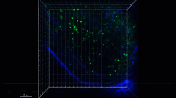
Now, NIH-funded scientists, led by Ronald Evans of the Salk Institute, La Jolla, CA, have devised a possible workaround: human islet-like organoids (HILOs) [1]. These tiny replicas of pancreatic tissue are created in the laboratory, and you can see them above secreting insulin (green) in a lab dish. Remarkably, some of these HILOs have been outfitted with a Harry Potter-esque invisibility cloak to enable them to evade immune attack when transplanted into mice.
Over several years, Doug Melton’s lab at Harvard University, Cambridge, MA, has worked steadily to coax induced pluripotent stem (iPS) cells, which are made from adult skin or blood cells, to form miniature islet-like cells in a lab dish [2]. My own lab at NIH has also been seeing steady progress in this effort, working with collaborators at the New York Stem Cell Foundation.
Although several years ago researchers could get beta cells to make insulin, they wouldn’t secrete the hormone efficiently when transplanted into a living mouse. About four years ago, the Evans lab found a possible solution by uncovering a genetic switch called ERR-gamma that when flipped, powered up the engineered beta cells to respond continuously to glucose and release insulin [3].
In the latest study, Evans and his team developed a method to program HILOs in the lab to resemble actual islets. They did it by growing the insulin-producing cells alongside each other in a gelatinous, three-dimensional chamber. There, the cells combined to form organoid structures resembling the shape and contour of the islet cells seen in an actual 3D human pancreas. After they are switched on with a special recipe of growth factors and hormones, these activated HILOs secrete insulin when exposed to glucose. When transplanted into a living mouse, this process appears to operate just like human beta cells work inside a human pancreas.
Another major advance was the invisibility cloak. The Salk team borrowed the idea from cancer immunotherapy and a type of drug called a checkpoint inhibitor. These drugs harness the body’s own immune T cells to attack cancer. They start with the recognition that T cells display a protein on their surface called PD-1. When T cells interact with other cells in the body, PD-1 binds to a protein on the surface of those cells called PD-L1. This protein tells the T cells not to attack. Checkpoint inhibitors work by blocking the interaction of PD-1 and PD-L1, freeing up immune cells to fight cancer.
Reversing this logic for the pancreas, the Salk team engineered HILOs to express PD-L1 on their surface as a sign to the immune system not to attack. The researchers then transplanted these HILOs into diabetic mice that received no immunosuppressive drugs, as would normally be the case to prevent rejection of these human cells. Not only did the transplanted HILOs produce insulin in response to glucose spikes, they spurred no immune response.
So far, HILOs transplants have been used to treat diabetes for more than 50 days in diabetic mice. More research will be needed to see whether the organoids can function for even longer periods of time.
Still, this is exciting news, and provides an excellent example of how advances in one area of science can provide new possibilities for others. In this case, these insights provide fresh hope for a day when children and adults with type 1 diabetes can live long, healthy lives without the need for frequent insulin injections.
References:
[1] Immune-evasive human islet-like organoids ameliorate diabetes. [published online ahead of print, 2020 Aug 19]. Yoshihara E, O’Connor C, Gasser E, Wei Z, Oh TG, Tseng TW, Wang D, Cayabyab F, Dai Y, Yu RT, Liddle C, Atkins AR, Downes M, Evans RM. Nature. 2020 Aug 19. [Epub ahead of publication]
[2] Generation of Functional Human Pancreatic β Cells In Vitro. Pagliuca FW, Millman JR, Gürtler M, Segel M, Van Dervort A, Ryu JH, Peterson QP, Greiner D, Melton DA. Cell. 2014 Oct 9;159(2):428-39.
[3] ERRγ is required for the metabolic maturation of therapeutically functional glucose-responsive β cells. Yoshihara E, Wei Z, Lin CS, Fang S, Ahmadian M, Kida Y, Tseng T, Dai Y, Yu RT, Liddle C, Atkins AR, Downes M, Evans RM. Cell Metab. 2016 Apr 12; 23(4):622-634.
Links:
Type 1 Diabetes (National Institute of Diabetes and Digestive and Kidney Diseases/NIH)
Pancreatic Islet Transplantation (National Institute of Diabetes and Digestive and Kidney Diseases)
“The Nobel Prize in Physiology or Medicine 2012” for Induced Pluripotent Stem Cells, The Nobel Prize news release, October 8, 2012.
Evans Lab (Salk Institute, La Jolla, CA)
NIH Support: National Institute of Diabetes and Digestive and Kidney Diseases; National Cancer Institute
First Virtual WALS Lecture
Posted on by Dr. Francis Collins

New Target for Cancer Immunotherapy: Exosomes
Posted on by Dr. Francis Collins

It was once a central tenet of biology that RNA molecules did their work inside the cell. But it’s now clear that RNA molecules are also active outside the cell, with potentially major implications for our health. To learn more about these unrecognized roles, the NIH Common Fund has launched the Extracellular RNA (exRNA) Communication Program.
This month, members of this research consortium described their latest progress in unraveling the secrets of exRNA in a group of 18 papers in the Cell family of journals. And it’s not just RNA that the consortium is studying, it’s also proteins. Among the many exciting results just published is the serendipitous discovery that proteins carried inside tiny, bubble-like vesicles, called exosomes, may influence a cancer’s response to immunotherapy [1]. The work sheds light on why certain cancers are resistant to immunotherapy and points to new strategies for unleashing the immune system in the fight against cancer.
The new findings center on a type of immunotherapy drugs known as checkpoint inhibitors. They are monoclonal antibodies produced by industry that can boost the immune system’s ability to attack and treat cancer.
One of those antibodies specifically targets a protein, called PD-1, on the surface of certain immune cells. When PD-1 binds a similarly named protein, called PD-L1, on the surface of another cell, the interaction prevents immune cells from attacking. Some tumors seem to have learned this and load up on PD-L1 to evade the immune system.
That’s where checkpoint inhibitors come in. By blocking the interaction between PD-1 and PD-L1, the treatment removes a key check on the immune system, allowing certain immune cells to wake up and attack the tumor.
Checkpoint inhibitors work better in some cancer types than in others. In melanoma, for example, up to about 30 percent of patients respond to checkpoint inhibitor therapy. But in prostate cancer, response rates are in the single digits.
Researchers led by Robert Blelloch, a member of the exRNA consortium and a scientist at the University of California, San Francisco, wanted to know why. He and his team looked for clues in RNA within the cells taken from immunotherapy-resistant prostate cancers.
As published in Cell, the researchers got their first hint of something biologically intriguing in an apparent discrepancy in their data. As they expected from prior work, PD-L1 protein was present in the treatment-resistant cancers. But the PD-L1 messenger RNAs (mRNA), which serve as templates for producing the protein, told an unexpected story. The resistant cancer cells made far more PD-L1 mRNAs than needed to produce the modest levels of PD-L1 proteins detected inside the cells.
Where was the missing PD-L1? Blelloch’s team found it in exosomes. The cancer cells were packaging large quantities of the protein inside exosomes and secreting them out of the cell to other parts of the body.
In additional studies with a mouse model of prostate cancer, the researchers found that those PD-L1-packed exosomes travel through the blood and lymphatic systems to lymph nodes, the sites where immune cells become activated. Once there, PD-L1-laden exosomes put the immune system to sleep, preventing certain key cells from locating and attacking the cancer, including the primary tumor and places where it may have spread.
In important follow up studies, the researchers edited two genes in cancer cells to prevent them from producing exosomes. And, in the absence of exosomes, the cells no longer formed tumors. Importantly, both edited and unedited cells still produced PD-L1, but only those that exported PD-L1 in exosomes disarmed the immune system. Studies in a mouse model of immunotherapy-resistant colorectal cancer yielded similar results.
The new evidence suggests that blocking the release of PD-L1 in exosomes, even temporarily, might allow the immune system to launch a successful and sustained attack against a cancer.
Blelloch notes that many intriguing questions remain. For example, it’s not yet clear why antibodies that target PD-L1 on cancer cells don’t disable PD-L1 found in exosomes. The good news is that the new findings suggest it may be possible to find small molecules that do target PD-L1-packed exosomes, unleashing the immune system against cancers that don’t respond to existing checkpoint inhibitors. In fact, Blelloch’s team is already screening for small molecules that might fit the bill.
Since its launch about five years ago, the exRNA Communication Program has published an impressive 480 peer-reviewed papers, including the latest work in the Cell family of journals. I’d encourage readers to click on some of the other excellent work. I hear that another batch of papers will be published later this year.
Reference:
[1] Suppression of exosomal PD-L induces systemic anti-tumor immunity and memory. Poggio M, Hu T, Pai CC, Chu B, Belair CD, Chang A, Montabana E, Lang UE, Fu Q, Fong L, Blelloch R. Cell. 2019 Apr 4;177(2):414-427.
Links:
Video: Unlocking the Mysteries of RNA Communication (Common Fund/NIH)
Immunotherapy to Treat Cancer (National Cancer Institute/NIH)
Blelloch Lab (University of California, San Francisco)
NIH Support: Common Fund; National Cancer Institute; National Center for Advancing Translational Sciences; National Heart, Lung, and Blood Institute; National Institute on Drug Abuse
Fighting Cancer with Natural Killer Cells
Posted on by Dr. Francis Collins
Credit: Michele Ardolino, University of Ottawa, and Brian Weist, Gilead Sciences, Foster City, CA
Cancer immunotherapies, which enlist a patient’s own immune system to attack and shrink developing tumors, have come a long way in recent years, leading in some instances to dramatic cures of widely disseminated cancers. But, as this video highlights, new insights from immunology are still being revealed that may provide even greater therapeutic potential.
Our immune system comes equipped with all kinds of specialized cells, including the infection-controlling Natural Killer (NK) cells. The video shows an army of NK cells (green) attacking a tumor in a mouse (blood vessels, blue) treated with a well-established type of cancer immunotherapy known as a checkpoint inhibitor. What makes the video so interesting is that researchers didn’t think checkpoint inhibitors could activate NK cells.
FDA Approves First CAR-T Cell Therapy for Pediatric Acute Lymphoblastic Leukemia
Posted on by Dr. Francis Collins

Credit: Emily Whitehead Foundation
Tremendous progress continues to be made against the Emperor of All Maladies, cancer. One of the most exciting areas of progress involves immunotherapy, a treatment strategy that harnesses the natural ability of the body’s own immune cells to attack and kill tumor cells. A lot of extremely hard work has gone into this research, so I was thrilled to learn that the Food and Drug Administration (FDA) just announced today its first approval of a promising type of immunotherapy called CAR-T cell therapy for kids and young adults with B-cell acute lymphoblastic leukemia (ALL)—the most common childhood cancer in the U.S.
ALL is a cancer of white blood cells called lymphocytes. Its treatment with chemotherapy drugs, developed with NIH support, has transformed ALL’s prognosis in kids from often fatal to largely treatable: about 90 percent of young patients now recover. But for those for whom the treatment fails, the prognosis is grim.
In the spring of 2012, Emily Whitehead of Philipsburg, PA was one such patient. The little girl was deathly ill, and her parents were worried they’d run out of options. That’s when doctors at Children’s Hospital of Philadelphia gave Emily and her parents new hope. Carl June and his team had successfully treated three adults with their version of CAR-T cell therapy, which is grounded in initial basic research supported by NIH [1,2]. Moving forward with additional clinical tests, they treated Emily—their first pediatric patient—that April. For a while, it was touch and go, and Emily almost died. But by May 2012, her cancer was in remission. Today, five years later, 12-year-old Emily remains cancer free and is thriving. And I’ve had the great privilege of getting to know Emily and her parents over the last few years.