A few years ago, I highlighted a really cool technology called Drop-seq for simultaneously analyzing the gene expression activity inside thousands of individual cells. Today, one of its creators, Evan Macosko, reports significant progress in developing even better tools for single-cell analysis—with support from an NIH Director’s New Innovator Award.
In a paper in the journal Science, Macosko, Fei Chen, and colleagues at the Broad Institute of Harvard and Massachusetts Institute of Technology (MIT), Cambridge, recently unveiled another exciting creation called Slide-seq [1]. This technology acts as a GPS-like system for mapping the exact location of each of the thousands of individual cells undergoing genomic analysis in a tissue sample.
This 3D video shows the exquisite precision of this new cellular form of GPS, which was used to generate a high-resolution map of the different cell types found in a tiny cube of mouse brain tissue. Specifically, it provides locations of the cell types and gene expression in the hippocampal regions called CA1 (green), CA2/3 (blue), and dentate gyrus (red).
Because using Slide-seq in the lab requires no specialized imaging equipment or skills, it should prove valuable to researchers across many different biomedical disciplines who want to look at cellular relationships or study gene activity in tissues, organs, or even whole organisms.
How does Slide-seq work? Macosko says one of the main innovations is an inexpensive rubber-coated glass slide nicknamed a puck. About 3 millimeters in diameter, pucks are studded with tens of thousands of 10 micron-sized beads, each one decorated with a random snippet of genetic material—an RNA barcode—that serves as its unique identifier of the bead.
The barcodes are sequenced en masse, and the exact location of each barcoded bead is indexed using innovative software developed by a team led by Chen, who is an NIH Director’s Early Independence awardee.
Then, the researchers place a sample of fresh-frozen tissue (typically, 10 micrometers, or 0.00039 inches, thick) on the puck and dissolve the tissue, lysing the cells and releasing their messenger RNA (mRNA). That leaves only the barcoded beads binding the mRNA transcripts expressed by the cells in the tissue—a biological record of the genes that were turned on at the time the sample was frozen.
The barcoded mRNA is then sequenced. The spatial position of each mRNA molecule can be inferred, using the reference index on the puck. This gives researchers a great deal of biological information about the cells in the tissue, often including their cell type and their gene expression pattern. All the data can then be mapped out in ways similar to those seen in this video, which was created using data from 66 pucks.
Slide-seq has been tested on a range of tissues from both mouse and human, replicating results from similar maps created using existing approaches, but also uncovering new biology. For example, in the mouse cerebellum, Slide-seq allowed the researchers to detect bands of variable gene activity across the tissues. This intriguing finding suggests that there may be subpopulations of cells in this part of the brain that have gene activity influenced by their physical locations.
Such results demonstrate the value of combining cell location with genomic information. In fact, Macosko now hopes to use Slide-seq to study the response of brain cells that are located near the buildup of damaged amyloid protein associated with the early-stage Alzheimer’s disease. Meanwhile, Chen is interested in pursuing cell lineage studies in a variety of tissues to see how and where changes in the molecular dynamics of tissues can lead to disease.
These are just a few examples of how Slide-seq will add to the investigative power of single-cell analysis in the years ahead. In meantime, the Macosko and Chen labs are working hard to develop even more innovative approaches to this rapidly emerging areas of biomedical research, so who knows what “seq” we will be talking about next?
Happy New Year! While everyone was busy getting ready for the holidays, the journal Science announced its annual compendium of scientific Breakthroughs of the Year. If you missed it, the winner for 2016 was the detection of gravitational waves—tiny ripples in the fabric of spacetime created by the collision of two black holes 1.3 billion years ago! It’s an incredible discovery, and one that Albert Einstein predicted a century ago.
Among the nine other advances that made the first cut for Breakthrough of the Year, several involved the biomedical sciences. As I’ve done in previous years (here and here), I’ll kick off this New Year by taking a quick look of some of the breakthroughs that directly involved NIH support:
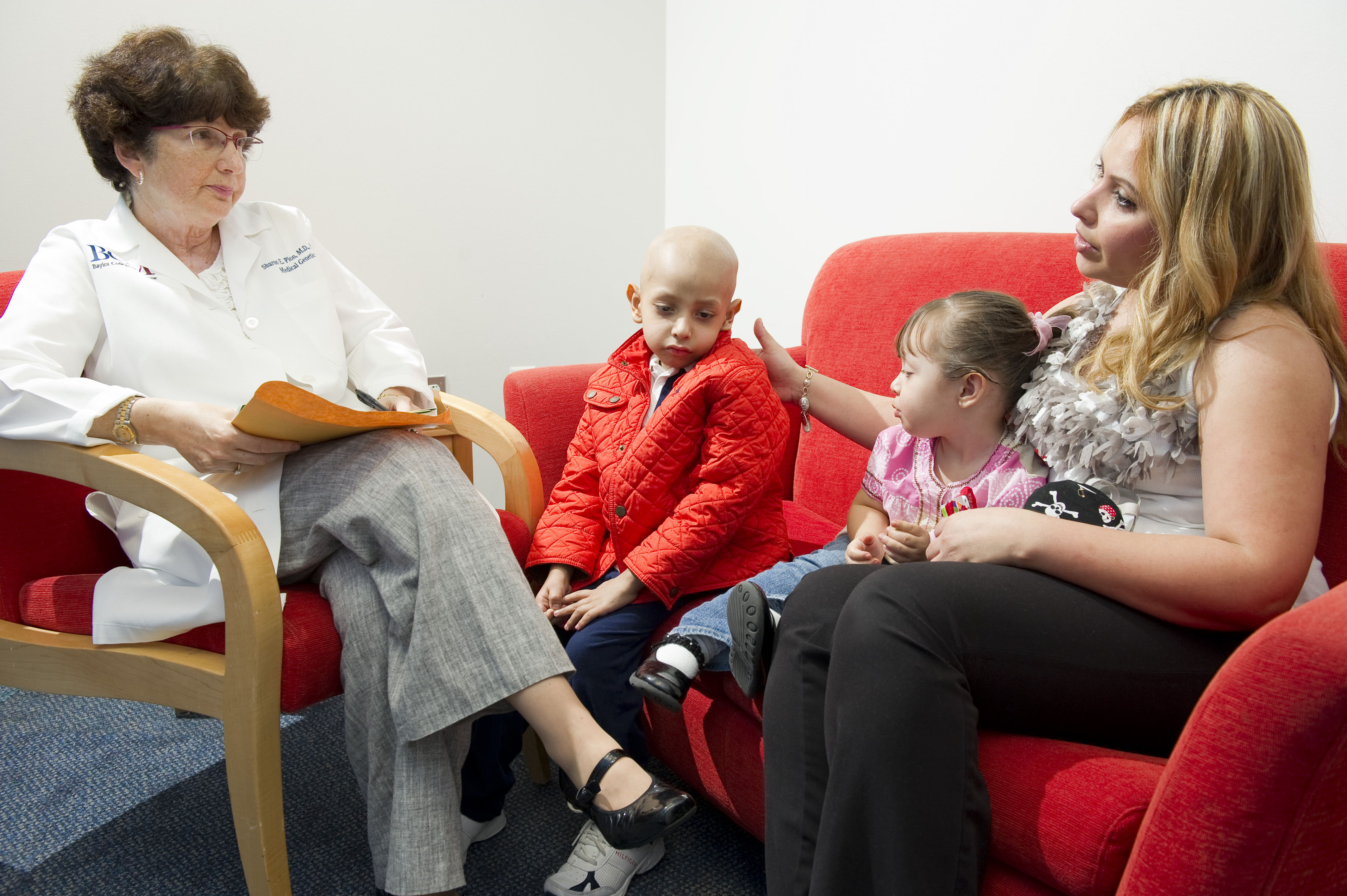
Caption: Baylor’s Sharon Plon consults with a family at the Texas Children’s Cancer Center in Houston. Credit: Paul V. Kuntz/Texas Children’s Hospital
An impressive number of fundamental advances in our understanding of cancer have occurred over the past several decades. One of the most profound is the realization that cancer is a disease of the genome, driven by a wide array of changes in DNA—some in the germline and affecting all cells of the body, but most occurring in individual cells during life (so-called “somatic mutations”). As the technology for sequencing cancer genomes has advanced, we are learning that virtually all cancers carry a unique set of mutations. Most are DNA copying errors of no significance (we call those “passengers”), but a few of them occur in genes that regulate cell growth and contribute causatively to the cancer (we call those “drivers”). We are now learning that it may be far more important for treating cancer to figure out what driver mutations are present in a patient’s tumor than to identify in which organ it arose. And, as a new study shows, this approach even appears to have potential to help cancer’s littlest victims.
Using genomic technology to analyze both tumor and blood samples from a large number of children who’d been newly diagnosed with cancer, an NIH-funded research team uncovered genetic clues with the potential to refine diagnosis, identify inherited cancer susceptibility, or guide treatment for nearly 40 percent of the children [1]. The potential driver mutations spanned a broad spectrum of genes previously implicated not only in pediatric cancers, but also in adult cancers. While much more work remains to determine how genomic analyses can be used to devise precise, new strategies for treating kids with cancer, the study provides an excellent example of the kind of research that NIH hopes to accelerate under the nation’s new cancer “moonshot,” a research initiative recently announced by the President and being led by the Vice President.
 Happy New Year! While everyone was busy getting ready for the holidays, the journal Science announced its annual compendium of scientific Breakthroughs of the Year. If you missed it, the winner for 2016 was the detection of gravitational waves—tiny ripples in the fabric of spacetime created by the collision of two black holes 1.3 billion years ago! It’s an incredible discovery, and one that Albert Einstein predicted a century ago.