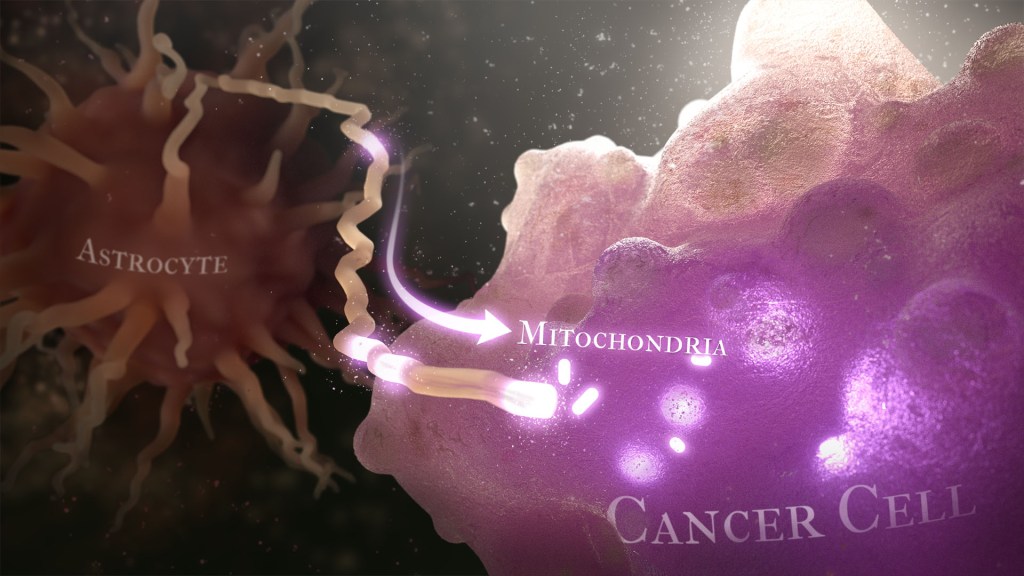
Caption: Illustration of cancer cell (bottom right) stealing mitochondria (white ovals) from a healthy astrocyte cell (left). Credit: Donny Bliss/NIH
Over the years, cancer researchers have uncovered many of the tricks that tumors use to fuel their growth and evade detection by the body’s immune system. More tricks await discovery, and finding them will be key in learning to target the right treatments to the right cancers.
Recently, a team of researchers demonstrated in lab studies a surprising trick pulled off by cells from a common form of brain cancer called glioblastoma. The researchers found that glioblastoma cells steal mitochondria, the power plants of our cells, from other cells in the central nervous system [1].
Why would cancer cells do this? How do they pull it off? The researchers don’t have all the answers yet. But glioblastoma arises from abnormal astrocytes, a particular type of the glial cell, a common cell in the brain and spinal cord. It seems from their initial work that stealing mitochondria from neighboring normal cells help these transformed glioblastoma cells to ramp up their growth. This trick might also help to explain why glioblastoma is one of the most aggressive forms of primary brain cancer, with limited treatment options.
In the new study, published in the journal Nature Cancer, a team co-led by Justin Lathia, Lerner Research Institute, Cleveland Clinic, OH, and Hrvoje Miletic, University of Bergen, Norway, had noticed some earlier studies suggesting that glioblastoma cells might steal mitochondria. They wanted to take a closer look.
This very notion highlights an emerging and much more dynamic view of mitochondria. Scientists used to think that mitochondria—which can number in the thousands within a single cell—generally just stayed put. But recent research has established that mitochondria can move around within a cell. They sometimes also get passed from one cell to another.
It also turns out that the intercellular movement of mitochondria has many implications for health. For instance, the transfer of mitochondria helps to rescue damaged tissues in the central nervous system, heart, and respiratory system. But, in other circumstances, this process may possibly come to the rescue of cancer cells.
While Lathia, Miletic, and team knew that mitochondrial transfer was possible, they didn’t know how relevant or dangerous it might be in brain cancers. To find out, they studied mice implanted with glioblastoma tumors from other mice or people with glioblastoma. This mouse model also had been modified to allow the researchers to trace the movement of mitochondria.
Their studies show that healthy cells often transfer some of their mitochondria to glioblastoma cells. They also determined that those mitochondria often came from healthy astrocytes, a process that had been seen before in the recovery from a stroke.
But the transfer process isn’t easy. It requires that a cell expend a lot of energy to form actin filaments that contract to pull the mitochondria along. They also found that the process depends on growth-associated protein 43 (GAP43), suggesting that future treatments aimed at this protein might help to thwart the process.
Their studies also show that, after acquiring extra mitochondria, glioblastoma cells shift into higher gear. The cancerous cells begin burning more energy as their metabolic pathways show increased activity. These changes allow for more rapid and aggressive growth. Overall, the findings show that this interaction between healthy and cancerous cells may partly explain why glioblastomas are so often hard to beat.
While more study is needed to confirm the role of this process in people with glioblastoma, the findings are an important reminder that treatment advances in oncology may come not only from study of the cancer itself but also by carefully considering the larger context and environments in which tumors grow. The hope is that these intriguing new findings will one day lead to new treatment options for the approximately 13,000 people in the U.S. alone who are diagnosed with glioblastoma each year [2].
NIH Support: National Institute of Neurological Disorders and Stroke; National Center for Advancing Translational Sciences; National Cancer Institute; National Institute of Allergy and Infectious Diseases
Most of the “cool” videos shared on my blog are borne of countless hours behind a microscope. Researchers must move a biological sample through a microscope’s focus, slowly acquiring hundreds of high-res 2D snapshots, one painstaking snap at a time. Afterwards, sophisticated computer software takes this ordered “stack” of images, calculates how the object would look from different perspectives, and later displays them as 3D views of life that can be streamed as short videos.
But this video is different. It was created by what’s called a multi-angle projection imaging system. This new optical device requires just a few camera snapshots and two mirrors to image a biological sample from multiple angles at once. Because the device eliminates the time-consuming process of acquiring individual image slices, it’s up to 100 times faster than current technologies and doesn’t require computer software to construct the movie. The kicker is that the video can be displayed in real time, which isn’t possible with existing image-stacking methods.
The video here shows two human melanoma cells, rotating several times between overhead and side views. You can see large amounts of the protein PI3K (brighter orange hues indicate higher concentrations), which helps some cancer cells divide and move around. Near the cell’s perimeter are small, dynamic surface protrusions. PI3K in these “blebs” is thought to help tumor cells navigate and survive in foreign tissues as the tumor spreads to other organs, a process known as metastasis.
The new multi-angle projection imaging system optical device was described in a paper published recently in the journal Nature Methods [1]. It was created by Reto Fiolka and Kevin Dean at the University of Texas Southwestern Medical Center, Dallas.
Like most technology, this device is complicated. Rather than the microscope and camera doing all the work, as is customary, two mirrors within the microscope play a starring role. During a camera exposure, these mirrors rotate ever so slightly and warp the acquired image in such a way that successive, unique perspectives of the sample magically come into view. By changing the amount of warp, the sample appears to rotate in real-time. As such, each view shown in the video requires only one camera snapshot, instead of acquiring hundreds of slices in a conventional scheme.
The concept traces to computer science and an algorithm called the shear warp transform method. It’s used to observe 3D objects from different perspectives on a 2D computer monitor. Fiolka, Dean, and team found they could implement a similar algorithm optically for use with a microscope. What’s more, their multi-angle projection imaging system is easy-to-use, inexpensive, and can be converted for use on any camera-based microscope.
The researchers have used the device to view samples spanning a range of sizes: from mitochondria and other tiny organelles inside cells to the beating heart of a young zebrafish. And, as the video shows, it has been applied to study cancer and other human diseases.
In a neat, but also scientifically valuable twist, the new optical method can generate a virtual reality view of a sample. Any microscope user wearing the appropriately colored 3D glasses immediately sees the objects.
While virtual reality viewing of cellular life might sound like a gimmick, Fiolka and Dean believe that it will help researchers use their current microscopes to see any sample in 3D—offering the chance to find rare and potentially important biological events much faster than is possible with even the most advanced microscopes today.
Fiolka, Dean, and team are still just getting started. Because the method analyzes tissue very quickly within a single image frame, they say it will enable scientists to observe the fastest events in biology, such as the movement of calcium throughout a neuron—or even a whole bundle of neurons at once. For neuroscientists trying to understand the brain, that’s a movie they will really want to see.