Seeing Coronavirus Replicate in Kidney Cells
Posted on by Dr. Francis Collins
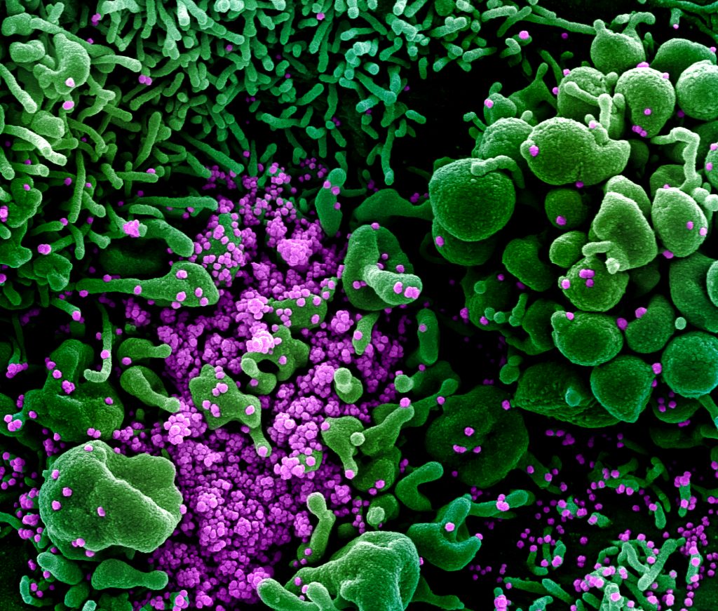
You’ve probably seen pictures of SARS-CoV-2—the novel coronavirus that causes COVID-19—that look alarming. But the high-resolution micrograph above paints a rather different picture, using rich pseudo-colors to show how newly assembled viral particles cause infected cells to bulge, or bleb, and then self-destruct.
This image depicts a common primate kidney cell line (green) infected with SARS-CoV-2. Notice the bulging, spherical cellular blebs, seen best in the upper right and bottom left corners. These badly damaged cells, which are filled to the point of bursting with viral particles, are beginning to self-destruct. Some cells have apparently already burst open, allowing hundreds of viral particles (purple) to spill out and potentially infect other cells.
This stunning picture was taken by John Bernbaum, an electron microscopist with NIH’s National Institute of Allergy and Infectious Diseases (NIAID). Bernbaum works at NIAID’s Integrated Research Facility (IRF), Fort Detrick, MD, a specialized, high-level biocontainment facility equipped with unique medical imaging capabilities. In this special environment, Bernbaum and his colleagues can safely visualize SARS-CoV-2, as well as other viruses and microbes that pose serious risks to human health.
To get this shot of SARS-CoV-2, Bernbaum relied on a conventional scanning electron microscope (SEM). First, a sample of kidney cells that had been exposed to SARS-CoV-2 was dehydrated, chemically preserved, and coated with a thin layer of metal. Once everything was ready, the SEM was used to focus a high-energy beam of electrons onto the sample. As electrons bounced off the metal surface, they revealed spatial variations and properties in the sample that were used to generate this 3D image.
Originally, this image was in gray scale. To better highlight the destructive powers of SARS-CoV-2, Jiro Wada, a skilled graphic illustrator at the IRF, used a computer program to colorize key features in exquisite detail. By studying these 3D images, researchers can learn about things such as the rate of infection and the prodigious number of particles each infected cell produces. They can also learn about how the infection affects the conditions inside cells.
Interestingly, what Bernbaum finds most striking about SARS-CoV-2 is what you don’t see in his images. Uninfected kidney cells look like a flat, delicately interwoven quilt (not pictured). When Bernbaum used SEM to study this sample of kidney cells, about 80 to 90 percent of the cells appeared flat and unremarkable. Yet, as the scan progressed, he came across a small subset of cells that appeared to be deformed by SARS-CoV-2 infection. Those abnormalities include the spherical bulges that I pointed out earlier, along with some worm-like protrusions that you can see in the top left.
Bernbaum has been producing amazing images like this one for 32 years—the last 11 of them at the IRF. If you’d like to see even more of his impressive work and that of the IRF team, check out the NIAID’s image gallery.
Links:
Coronavirus (NIH)
Integrated Research Facility (National Institute of Allergy and Infectious Diseases/NIH)
NIH Support: National Institute of Allergy and Infectious Diseases
Beautiful and important work!
You could profit if you now look at photos of cells previously infected with Hep. B when they encounter Hep. D since this indicative of a Corona Virus attack. Massive geometric disruption leading to organ failure.
Thank you for the amazing shot. Question: How does the dehydration process affect the resulting image/primate cells, the morphology?
Mind blowing after you read and look at the scientific images provided. Science truly provides beautiful and alarming truths. Thank you for your important work and contribution.
And now we know why all of CKD ended up on the high risk of severe outcome.
Awesome to see it in such vivid color! Art in Medicine lives!
Kaleidoscopic visual elegant display of primate kidney cells with SARS-Covid19 depiction for overall scientific advanced intellectual insights in the complex immunopharmacology area!
Why don’t we not stimulate the production of GLUTATHIONE, our body’s antioxidant to boost our immune system?
A small protein produced naturally in the cells of our body, provides crucial protective functions.
Glutathione is our body’s protection from bacteria, fungus, viruses and other antigens. It is critical for healthy immune function. Glutathione can boost our immune system, help raise the antibodies to neutralize viruses like Covid-19.
Vaccines can certainly help in its role to raise antibodies, but it takes enormous time to develop, produce and have great uncertainty in its level of effectiveness.
I think we are missing out on something. There are over 100,000 articles on Glutathione on PubMed National Library of Medicine.
Can’t wait for the images of the virus causing cell death in human cell line. Come on, it’s been 18 months.