


Most of the “cool” videos shared on my blog are borne of countless hours behind a microscope. Researchers must move a biological sample through a microscope’s focus, slowly acquiring hundreds of high-res 2D snapshots, one painstaking snap at a time. Afterwards, sophisticated computer software takes this ordered “stack” of images, calculates how the object would look from different perspectives, and later displays them as 3D views of life that can be streamed as short videos.
But this video is different. It was created by what’s called a multi-angle projection imaging system. This new optical device requires just a few camera snapshots and two mirrors to image a biological sample from multiple angles at once. Because the device eliminates the time-consuming process of acquiring individual image slices, it’s up to 100 times faster than current technologies and doesn’t require computer software to construct the movie. The kicker is that the video can be displayed in real time, which isn’t possible with existing image-stacking methods.
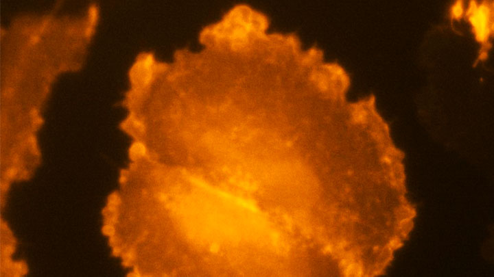
The video here shows two human melanoma cells, rotating several times between overhead and side views. You can see large amounts of the protein PI3K (brighter orange hues indicate higher concentrations), which helps some cancer cells divide and move around. Near the cell’s perimeter are small, dynamic surface protrusions. PI3K in these “blebs” is thought to help tumor cells navigate and survive in foreign tissues as the tumor spreads to other organs, a process known as metastasis.
The new multi-angle projection imaging system optical device was described in a paper published recently in the journal Nature Methods [1]. It was created by Reto Fiolka and Kevin Dean at the University of Texas Southwestern Medical Center, Dallas.
Like most technology, this device is complicated. Rather than the microscope and camera doing all the work, as is customary, two mirrors within the microscope play a starring role. During a camera exposure, these mirrors rotate ever so slightly and warp the acquired image in such a way that successive, unique perspectives of the sample magically come into view. By changing the amount of warp, the sample appears to rotate in real-time. As such, each view shown in the video requires only one camera snapshot, instead of acquiring hundreds of slices in a conventional scheme.
The concept traces to computer science and an algorithm called the shear warp transform method. It’s used to observe 3D objects from different perspectives on a 2D computer monitor. Fiolka, Dean, and team found they could implement a similar algorithm optically for use with a microscope. What’s more, their multi-angle projection imaging system is easy-to-use, inexpensive, and can be converted for use on any camera-based microscope.
The researchers have used the device to view samples spanning a range of sizes: from mitochondria and other tiny organelles inside cells to the beating heart of a young zebrafish. And, as the video shows, it has been applied to study cancer and other human diseases.
In a neat, but also scientifically valuable twist, the new optical method can generate a virtual reality view of a sample. Any microscope user wearing the appropriately colored 3D glasses immediately sees the objects.
While virtual reality viewing of cellular life might sound like a gimmick, Fiolka and Dean believe that it will help researchers use their current microscopes to see any sample in 3D—offering the chance to find rare and potentially important biological events much faster than is possible with even the most advanced microscopes today.
Fiolka, Dean, and team are still just getting started. Because the method analyzes tissue very quickly within a single image frame, they say it will enable scientists to observe the fastest events in biology, such as the movement of calcium throughout a neuron—or even a whole bundle of neurons at once. For neuroscientists trying to understand the brain, that’s a movie they will really want to see.
Reference:
[1] Real-time multi-angle projection imaging of biological dynamics. Chang BJ, Manton JD, Sapoznik E, Pohlkamp T, Terrones TS, Welf ES, Murali VS, Roudot P, Hake K, Whitehead L, York AG, Dean KM, Fiolka R. Nat Methods. 2021 Jul;18(7):829-834.
Links:
Metastatic Cancer: When Cancer Spreads (National Cancer Institute)
Fiolka Lab (University of Texas Southwestern Medical Center, Dallas)
Dean Lab (University of Texas Southwestern)
Microscopy Innovation Lab (University of Texas Southwestern)
NIH Support: National Cancer Institute; National Institute of General Medical Sciences