X-Ray Diffraction: Still Beautiful After All These Years
Posted on by Dr. Francis Collins
Source: John Johnson, The Scripps Research Institute
This year marks the 100th anniversary of X-ray diffraction technology. Developed in 1912, this important tool enables researchers to figure out the 3-D structure of a molecule by beaming X-rays, often through its crystallized form. More than 85% of the protein structures we know today have been determined via this method.
For more information about x-ray diffraction, I recommend Structural Biology Fact Sheet and The Structures of Life: X-ray Crystallography.

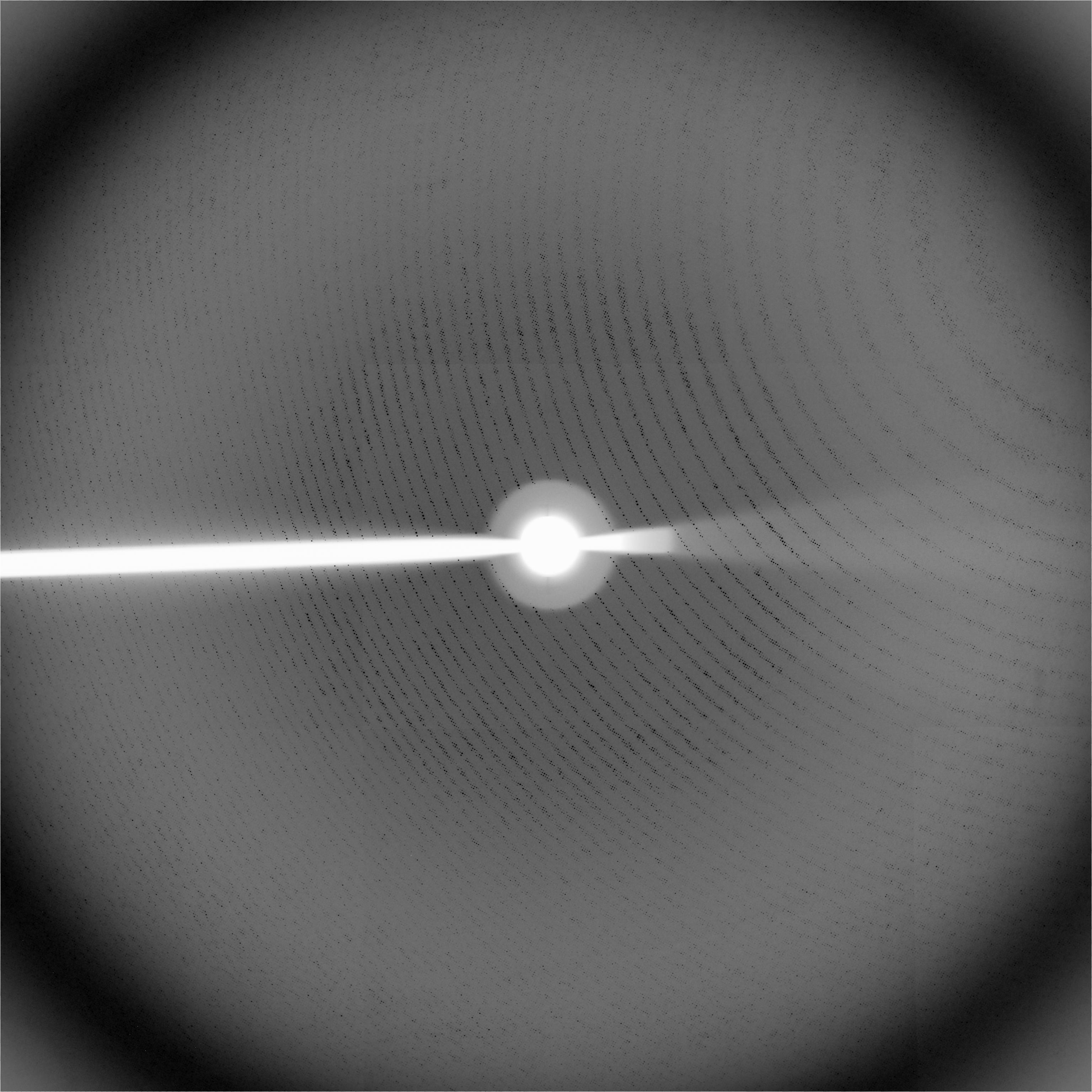
Here you see the X-ray diffraction image that James Watson and Francis Crick used to decipher the double helix structure of DNA in 1953.
And now for a trivia question! As some of you may know, one of my hobbies is playing the guitar—a guitar that happens to have a DNA double helix inlaid on its fretboard. All special guitars should have a name. B.B. King has Lucille. Eric Clapton had Blackie. After which famous scientist, responsible for the image used by Watson and Crick, is my guitar named?