light microscopy
Cool Videos: Pushing the Limits of Live-Cell Microscopy
Posted on by Dr. Francis Collins

If you’re not watching recent work in biology, you might have thought that light microscopy hit its limits years ago. After all, it’s been around a long time. But to the contrary, microscopic imaging technology just keeps getting better and better. Here you can look with unprecedented clarity at just one of the many dynamic processes going on within a living cell. Specifically, this video shows actin fibers (orange-red), which are key components of the cell’s cytoskeleton, slowly pulling clathrin-coated pits (green), which are basket-like structures containing molecular cargo, away from the cell’s external membrane and deeper within the cell.
This remarkable live-action view was produced using one of two new forms of extended-resolution, structured illumination microscopy (SIM). SIM is faster than other forms of super-resolution fluorescence microscopy. It’s also less damaging to cells, making it the go-to method for live-cell imaging. The downside has been SIM’s limited resolution—just twice that of conventional light microscopes. However, Nobel Prize-winner Eric Betzig and postdoc Dong Li of Howard Hughes Medical Institute, Janelia Research Campus, Ashburn, VA, along with colleagues including Jordan Beach and John Hammer at NIH’s National Heart, Lung, and Blood Institute, recently came up with two different solutions to enhance SIM’s spatial resolution.
LabTV: Curious about Microscopy
Posted on by Dr. Francis Collins
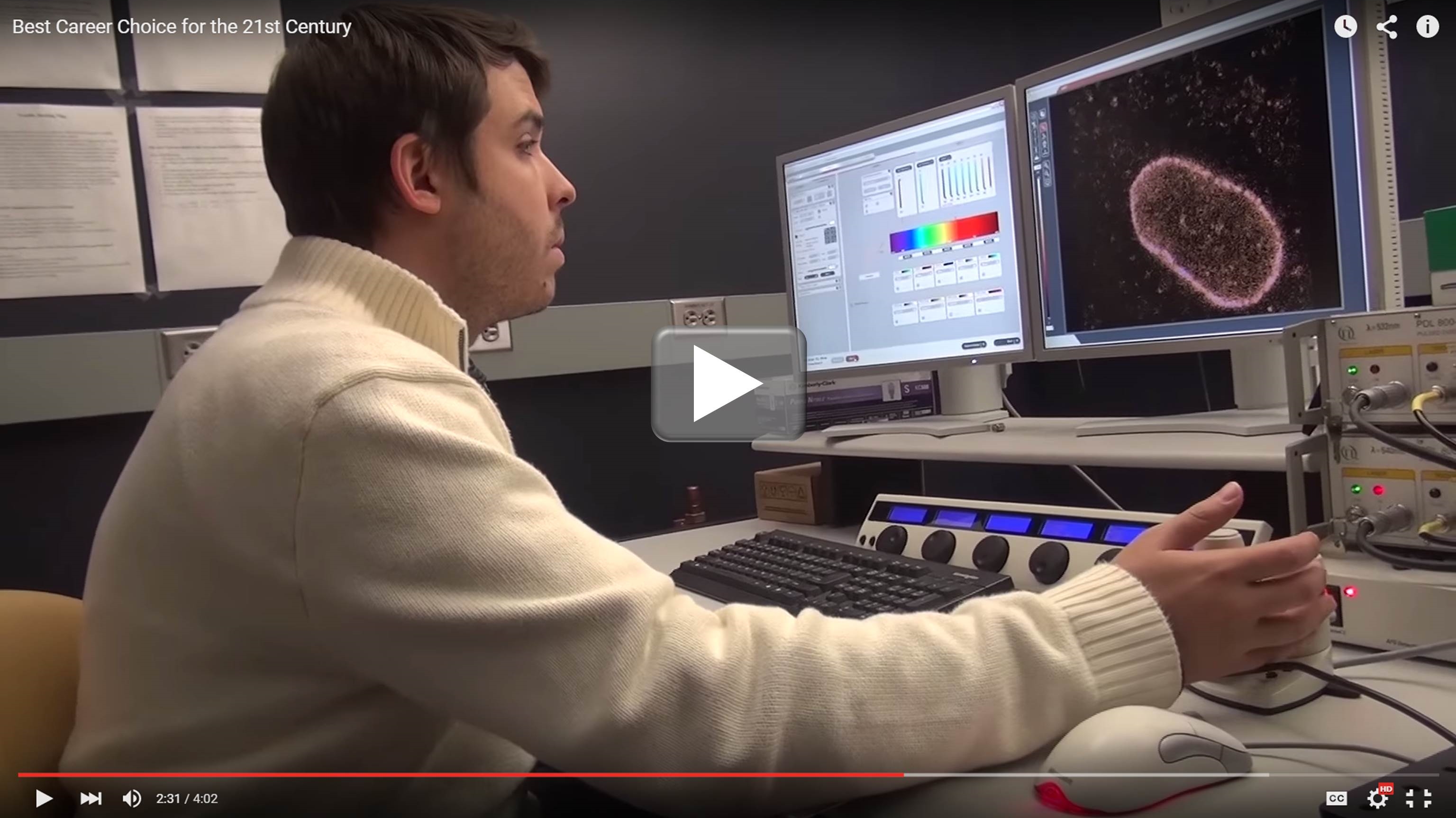
Growing up amid the potato and corn fields of western New York state, Jordan Myers got a firsthand look at what it was like to work as a farmer, a homebuilder, even a chimney sweep. But it was television—specifically, “Bill Nye the Science Guy” and “The Magic School Bus”—that introduced him to what would become his future career: science.
Propelled by his curiosity about how living things work, Myers left his hometown of Savannah to attend New York’s Rochester Institute of Technology, where he earned an undergraduate degree in biotechnology, and then headed off to pursue advanced degrees in cell biology at Yale School of Medicine, New Haven, CT. There, as you’ll see in this LabTV profile, he’s trying to develop light microscopy techniques [1,2] to view the cell’s nuclear envelope at nanometer (nm) resolution—a major challenge when one considers that a red blood cell measures about 7,000 nm in diameter.