CFC
A Cardboard Memory
Posted on by Dr. Francis Collins

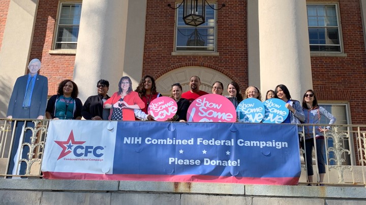
Kicking Off 2019 Combined Federal Campaign
Posted on by Dr. Francis Collins

Snapshots of Life: Lighting up the Promise of Retinal Gene Therapy
Posted on by Dr. Francis Collins

Caption: Large-scale mosaic confocal micrograph showing expression of a marker gene (yellow) transferred by gene therapy techniques into the ganglion cells (blue) of a mouse retina.
Credit: Keunyoung Kim, Wonkyu Ju, and Mark Ellisman, National Center for Microscopy and Imaging Research, University of California, San Diego
The retina, like this one from a mouse that is flattened out and captured in a beautiful image, is a thin tissue that lines the back of the eye. Although only about the size of a postage stamp, the retina contains more than 100 distinct cell types that are organized into multiple information-processing layers. These layers work together to absorb light and translate it into electrical signals that stream via the optic nerve to the brain.
In people with inherited disorders in which the retina degenerates, an altered gene somewhere within this nexus of cells progressively robs them of their sight. This has led to a number of human clinical trials—with some encouraging progress being reported for at least one condition, Leber congenital amaurosis—that are transferring a normal version of the affected gene into retinal cells in hopes of restoring lost vision.
To better understand and improve this potential therapeutic strategy, researchers are gauging the efficiency of gene transfer into the retina via an imaging technique called large-scale mosaic confocal microscopy, which computationally assembles many small, high-resolution images in a way similar to Google Earth. In the example you see above, NIH-supported researchers Wonkyu Ju, Mark Ellisman, and their colleagues at the University of California, San Diego, engineered adeno-associated virus serotype 2 (AAV2) to deliver a dummy gene tagged with a fluorescent marker (yellow) into the ganglion cells (blue) of a mouse retina. Two months after AAV-mediated gene delivery, yellow had overlaid most of the blue, indicating the dummy gene had been selectively transferred into retinal ganglion cells at a high rate of efficiency [1].
