CA3
Mapping the Brain’s Memory Bank
Posted on by Dr. Francis Collins
There’s a lot of groundbreaking research now underway to map the organization and internal wiring of the brain’s hippocampus, essential for memory, emotion, and spatial processing. This colorful video depicting a mouse hippocampus offers a perfect case in point.
The video presents the most detailed 3D atlas of the hippocampus ever produced, highlighting its five previously defined zones: dentate gyrus, CA1, CA2, CA3, and subiculum. The various colors within those zones represent areas with newly discovered and distinctive patterns of gene expression, revealing previously hidden layers of structural organization.
For instance, the subiculum, which sends messages from the hippocampus to other parts of the brain, includes several subregions. The subregions include the three marked in red, yellow, and blue at about 23 seconds into the video.
How’d the researchers do it? In the new study, published in Nature Neuroscience, the researchers started with the Allen Mouse Brain Atlas, a rich, publicly accessible 3D atlas of gene expression in the mouse brain. The team, led by Hong-Wei Dong, University of Southern California, Los Angeles, drilled down into the data to pull up 258 genes that are differentially expressed in the hippocampus and might be helpful for mapping purposes.
Some of those 258 genes were generally expressed only in previously defined portions of the hippocampus. Others were “turned on” only in discrete portions of known hippocampal domains, leading the researchers to define 20 distinct subregions that hadn’t been recognized before.
Combining these data, sophisticated analytical tools, and plenty of hard work, the team assembled this detailed atlas, together with connectivity data, to create a detailed wiring diagram. It includes about 200 signaling pathways that show how all those subregions network together and with other portions of the brain.
What’s really interesting is that the data also showed that these components of the hippocampus contribute to three relatively independent brain-wide communication networks. While much more study is needed, those three networks appear to relate to distinct functions of the hippocampus, including spatial navigation, social behaviors, and metabolism.
This more-detailed view of the hippocampus is just the latest from the NIH-funded Mouse Connectome Project. The ongoing project aims to create a complete connectivity atlas for the entire mouse brain.
The Mouse Connectome Project isn’t just for those with an interest in mice. Indeed, because the mouse and human brain are similarly organized, studies in the smaller mouse brain can help to provide a template for making sense of the larger and more complex human brain, with its tens of billions of interconnected neurons.
Ultimately, the hope is that this understanding of healthy brain connections will provide clues for better treating the brain’s abnormal connections and/or disconnections. They are involved in numerous neurological conditions, including Alzheimer’s disease, Parkinson’s disease, and autism spectrum disorder.
Reference:
[1] Integration of gene expression and brain-wide connectivity reveals the multiscale organization of mouse hippocampal networks. Bienkowski MS, Bowman I, Song MY, Gou L, Ard T, Cotter K, Zhu M, Benavidez NL, Yamashita S, Abu-Jaber J, Azam S, Lo D, Foster NN, Hintiryan H, Dong HW. Nat Neurosci. 2018 Nov;21(11):1628-1643.
Links:
Mouse Connectome Project (University of Southern California, Los Angeles)
Human Connectome Project (USC)
Allen Brain Map (Allen Institute, Seattle)
The Brain Research through Advancing Innovative Neurotechnologies® (BRAIN) Initiative (NIH)
NIH Support: National Institute of Mental Health; National Cancer Institute
Unlocking the Brain’s Memory Retrieval System
Posted on by Dr. Francis Collins

Credit:Sahay Lab, Massachusetts General Hospital, Boston
Play the first few bars of any widely known piece of music, be it The Star-Spangled Banner, Beethoven’s Fifth, or The Rolling Stones’ (I Can’t Get No) Satisfaction, and you’ll find that many folks can’t resist filling in the rest of the melody. That’s because the human brain thrives on completing familiar patterns. But, as we grow older, our pattern completion skills often become more error prone.
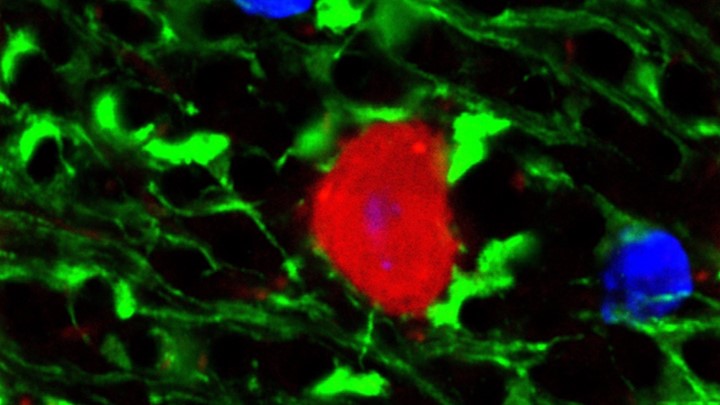
This image shows some of the neural wiring that controls pattern completion in the mammalian brain. Specifically, you’re looking at a cross-section of a mouse hippocampus that’s packed with dentate granule neurons and their signal-transmitting arms, called axons, (light green). Note how the axons’ short, finger-like projections, called filopodia (bright green), are interacting with a neuron (red) to form a “memory trace” network. Functioning much like an online search engine, memory traces use bits of incoming information, like the first few notes of a song, to locate and pull up more detailed information, like the complete song, from the brain’s repository of memories in the cerebral cortex.
Snapshots of Life: Color Coding the Hippocampus
Posted on by Dr. Francis Collins
The final frontier? Trekkies would probably say it’s space, but mapping the brain—the most complicated biological structure in the known universe—is turning out to be an amazing adventure in its own right. Not only are researchers getting better at charting the brain’s densely packed and varied cellular topography, they are starting to identify the molecules that neurons use to connect into the distinct information-processing circuits that allow all walks of life to think and experience the world.
This image shows distinct neural connections in a cross section of a mouse’s hippocampus, a region of the brain involved in the memory of facts and events. The large, crescent-shaped area in green is hippocampal zone CA1. Its highly specialized neurons, called place cells, serve as the brain’s GPS system to track location. It appears green because these neurons express cadherin-10. This protein serves as a kind of molecular glue that likely imparts specific functional properties to this region. [1]

