brain surgery
Artificial Intelligence Speeds Brain Tumor Diagnosis
Posted on by Dr. Francis Collins

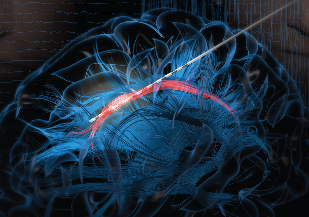
Credit: Joe Hallisy, Michigan Medicine, Ann Arbor
Computers are now being trained to “see” the patterns of disease often hidden in our cells and tissues. Now comes word of yet another remarkable use of computer-generated artificial intelligence (AI): swiftly providing neurosurgeons with valuable, real-time information about what type of brain tumor is present, while the patient is still on the operating table.
This latest advance comes from an NIH-funded clinical trial of 278 patients undergoing brain surgery. The researchers found they could take a small tumor biopsy during surgery, feed it into a trained computer in the operating room, and receive a diagnosis that rivals the accuracy of an expert pathologist.
Traditionally, sending out a biopsy to an expert pathologist and getting back a diagnosis optimally takes about 40 minutes. But the computer can do it in the operating room on average in under 3 minutes. The time saved helps to inform surgeons how to proceed with their delicate surgery and make immediate and potentially life-saving treatment decisions to assist their patients.
As reported in Nature Medicine, researchers led by Daniel Orringer, NYU Langone Health, New York, and Todd Hollon, University of Michigan, Ann Arbor, took advantage of AI and another technological advance called stimulated Raman histology (SRH). The latter is an emerging clinical imaging technique that makes it possible to generate detailed images of a tissue sample without the usual processing steps.
The SRH technique starts off by bouncing laser light rapidly through a tissue sample. This light enables a nearby fiberoptic microscope to capture the cellular and structural details within the sample. Remarkably, it does so by picking up on subtle differences in the way lipids, proteins, and nucleic acids vibrate when exposed to the light.
Then, using a virtual coloring program, the microscope quickly pieces together and colors in the fine structural details, pixel by pixel. The result: a high-resolution, detailed image that you might expect from a pathology lab, minus the staining of cells, mounting of slides, and the other time-consuming processing procedures.
To interpret the SRH images, the researchers turned to computers and machine learning. To teach a computer, it must be fed large datasets of examples in order to learn how to perform a given task. In this case, they used a special class of machine learning called deep neural networks, or deep learning. It’s inspired by the way neural networks in the human brain process information.
In deep learning, computers look for patterns in large collections of data. As they begin to recognize complex relationships, some connections in the network are strengthened while others are weakened. The finished network is typically composed of multiple information-processing layers, which operate on the data to return a result, in this case a brain tumor diagnosis.
The team trained the computer to classify tissues samples into one of 13 categories commonly found in a brain tumor sample. Those categories included the most common brain tumors: malignant glioma, lymphoma, metastatic tumors, and meningioma. The training was based on more than 2.5 million labeled images representing samples from 415 patients.
Next, they put the machine to the test. The researchers split each of 278 brain tissue samples into two specimens. One was sent to a conventional pathology lab for prepping and diagnosis. The other was imaged with SRH, and then the trained machine made a diagnosis.
Overall, the machine’s performance was quite impressive, returning the right answer about 95 percent of the time. That’s compared to an accuracy of 94 percent for conventional pathology.
Interestingly, the machine made a correct diagnosis in all 17 cases that a pathologist got wrong. Likewise, the pathologist got the right answer in all 14 cases in which the machine slipped up.
The findings show that the combination of SRH and AI can be used to make real-time predictions of a patient’s brain tumor diagnosis to inform surgical decision-making. That may be especially important in places where expert neuropathologists are hard to find.
Ultimately, the researchers suggest that AI may yield even more useful information about a tumor’s underlying molecular alterations, adding ever greater precision to the diagnosis. Similar approaches are also likely to work in supplying timely information to surgeons operating on patients with other cancers too, including cancers of the skin and breast. The research team has made a brief video to give you a more detailed look at the new automated tissue-to-diagnosis pipeline.
Reference:
[1] Near real-time intraoperative brain tumor diagnosis using stimulated Raman histology and deep neural networks. Hollon TC, Pandian B, Adapa AR, Urias E, Save AV, Khalsa SSS, Eichberg DG, D’Amico RS, Farooq ZU, Lewis S, Petridis PD, Marie T, Shah AH, Garton HJL, Maher CO, Heth JA, McKean EL, Sullivan SE, Hervey-Jumper SL, Patil PG, Thompson BG, Sagher O, McKhann GM 2nd, Komotar RJ, Ivan ME, Snuderl M, Otten ML, Johnson TD, Sisti MB, Bruce JN, Muraszko KM, Trautman J, Freudiger CW, Canoll P, Lee H, Camelo-Piragua S, Orringer DA. Nat Med. 2020 Jan 6.
Links:
Video: Artificial Intelligence: Collecting Data to Maximize Potential (NIH)
New Imaging Technique Allows Quick, Automated Analysis of Brain Tumor Tissue During Surgery (National Institute of Biomedical Imaging and Bioengineering/NIH)
Daniel Orringer (NYU Langone, Perlmutter Cancer Center, New York City)
Todd Hollon (University of Michigan, Ann Arbor)
NIH Support: National Cancer Institute; National Institute of Biomedical Imaging and Bioengineering
Discovering a Source of Laughter in the Brain
Posted on by Dr. Francis Collins
If laughter really is the best medicine, wouldn’t it be great if we could learn more about what goes on in the brain when we laugh? Neuroscientists recently made some major progress on this front by pinpointing a part of the brain that, when stimulated, never fails to induce smiles and laughter.
In their study conducted in three patients undergoing electrical stimulation brain mapping as part of epilepsy treatment, the NIH-funded team found that stimulation of a specific tract of neural fibers, called the cingulum bundle, triggered laughter, smiles, and a sense of calm. Not only do the findings shed new light on the biology of laughter, researchers hope they may also lead to new strategies for treating a range of conditions, including anxiety, depression, and chronic pain.
In people with epilepsy whose seizures are poorly controlled with medication, surgery to remove seizure-inducing brain tissue sometimes helps. People awaiting such surgeries must first undergo a procedure known as intracranial electroencephalography (iEEG). This involves temporarily placing 10 to 20 arrays of tiny electrodes in the brain for up to several weeks, in order to pinpoint the source of a patient’s seizures in the brain. With the patient’s permission, those electrodes can also enable physician-researchers to stimulate various regions of the patient’s brain to map their functions and make potentially new and unexpected discoveries.
In the new study, published in The Journal of Clinical Investigation, Jon T. Willie, Kelly Bijanki, and their colleagues at Emory University School of Medicine, Atlanta, looked at a 23-year-old undergoing iEEG for 8 weeks in preparation for surgery to treat her uncontrolled epilepsy [1]. One of the electrodes implanted in her brain was located within the cingulum bundle and, when that area was stimulated for research purposes, the woman experienced an uncontrollable urge to laugh. Not only was the woman given to smiles and giggles, she also reported feeling relaxed and calm.
As a further and more objective test of her mood, the researchers asked the woman to interpret the expression of faces on a computer screen as happy, sad, or neutral. Electrical stimulation to the cingulum bundle led her to see those faces as happier, a sign of a generally more positive mood. A full evaluation of her mental state also showed she was fully aware and alert.
To confirm the findings, the researchers looked to two other patients, a 40-year-old man and a 28-year-old woman, both undergoing iEEG in the course of epilepsy treatment. In those two volunteers, stimulation of the cingulum bundle also triggered laughter and reduced anxiety with otherwise normal cognition.
Willie notes that the cingulum bundle links many brain areas together. He likens it to a super highway with lots of on and off ramps. He suspects the spot they’ve uncovered lies at a key intersection, providing access to various brain networks regulating mood, emotion, and social interaction.
Previous research has shown that stimulation of other parts of the brain can also prompt patients to laugh. However, what makes stimulation of the cingulum bundle a particularly promising approach is that it not only triggers laughter, but also reduces anxiety.
The new findings suggest that stimulation of the cingulum bundle may be useful for calming patients’ anxieties during neurosurgeries in which they must remain awake. In fact, Willie’s team did so during their 23-year-old woman’s subsequent epilepsy surgery. Each time she became distressed, the stimulation provided immediate relief. Also, if traditional deep brain stimulation or less invasive means of brain stimulation can be developed and found to be safe for long-term use, they may offer new ways to treat depression, anxiety disorders, and/or chronic pain.
Meanwhile, Willie’s team is hard at work using similar approaches to map brain areas involved in other aspects of mood, including fear, sadness, and anxiety. Together with the multidisciplinary work being mounted by the NIH-led BRAIN Initiative, these kinds of studies promise to reveal functionalities of the human brain that have previously been out of reach, with profound consequences for neuroscience and human medicine.
Reference:
[1] Cingulum stimulation enhances positive affect and anxiolysis to facilitate awake craniotomy. Bijanki KR, Manns JR, Inman CS, Choi KS, Harati S, Pedersen NP, Drane DL, Waters AC, Fasano RE, Mayberg HS, Willie JT. J Clin Invest. 2018 Dec 27.
Links:
Video: Patient’s Response (Bijanki et al. The Journal of Clinical Investigation)
Epilepsy Information Page (National Institute of Neurological Disease and Stroke/NIH)
Jon T. Willie (Emory University, Atlanta, GA)
NIH Support: National Institute of Neurological Disease and Stroke; National Center for Advancing Translational Sciences
How the Brain Regulates Vocal Pitch
Posted on by Dr. Francis Collins
Credit: University of California, San Francisco
Whether it’s hitting a high note, delivering a punch line, or reading a bedtime story, the pitch of our voices is a vital part of human communication. Now, as part of their ongoing quest to produce a dynamic picture of neural function in real time, researchers funded by the NIH’s Brain Research through Advancing Innovative Neurotechnologies (BRAIN) Initiative have identified the part of the brain that controls vocal pitch [1].
This improved understanding of how the human brain regulates the pitch of sounds emanating from the voice box, or larynx, is more than cool neuroscience. It could aid in the development of new, more natural-sounding technologies to assist people who have speech disorders or who’ve had their larynxes removed due to injury or disease.