Past Stories
Turning Discoveries into Health for All
Posted on by Dr. Monica M. Bertagnolli

Greetings, blog readers! I’m Dr. Monica Bertagnolli, and I’m honored to be serving as the 17th Director of the National Institutes of Health. I’m excited to continue the NIH Director’s Blog to share with you the exciting discoveries and fascinating research conducted here at NIH and at the organizations we support in the U.S. and around the world. But before we start diving into the latest advances, I wanted to share a bit about myself and what I’m looking forward to as NIH Director.
I spent most of my career caring for people with cancer as a surgical oncologist and researcher before joining NIH last year as Director of the National Cancer Institute. While I miss the operating room (although I couldn’t stay away for long—more about that in a forthcoming blog!) and the opportunity to work with patients every day, I’m eager to serve the public in my new role as NIH Director.
When I was growing up, my family raised sheep and cattle on a ranch at the base of the Wind River Mountains in Wyoming. I know the health challenges that come with living in a rural area: Not everyone has access to an academic medical center and clinical studies, and managing the logistics of routine and preventive check-ups can be difficult. Unfortunately, many of our research advances are not reaching enough people in these areas.
As a cancer survivor, I am keenly aware that I’ve been fortunate to have access to excellent care, which has been directly informed by NIH-funded research over the past five decades. I know the transformative power of research to save lives, but from my experience as a clinician, I know that it is not always possible for people to receive the care that they need due to financial, geographic, or cultural barriers. It is unacceptable for the benefits of NIH-funded biomedical research to be available to some but not all.
That’s why one of my goals as NIH Director is to ensure the biomedical research enterprise and its discoveries—from basic to clinical research—are more inclusive and accessible to people from all walks of life, including rural areas. Income, age, race, ethnicity, geographic location, and disability status should not be barriers to participating in research or to benefitting from research advances. By meeting people where they are and engaging more communities as our research partners, I believe we can also make significant progress in rebuilding trust in science across the country.
Right now, we have an unprecedented opportunity to embrace and increase access to innovation: Our knowledge and technology have developed to the point that we should be able to deliver evidence-based, data-driven health care to everyone. This is an exciting time for science, and I can’t wait to share more with you in the weeks and months to come.
Experiencing the Neural Symphony Underlying Memory through a Blend of Science and Art
Posted on by John Ngai, PhD, NIH BRAIN Initiative
Ever wonder how you’re able to remember life events that happened days, months, or even years ago? You have your hippocampus to thank. This essential area in the brain relies on intense and highly synchronized patterns of activity that aren’t found anywhere else in the brain. They’re called “sharp-wave ripples.”
These dynamic ripples have been likened to the brain version of an instant replay, appearing most commonly during rest after a notable experience. And, now, the top video winner in this year’s Brain Research Through Advancing Innovative Neurotechnologies® (BRAIN) Initiative’s annual Show Us Your BRAINs! Photo and Video Contest allows you to witness the “chatter” that those ripples set off in other neurons. The details of this chatter determine just how durable a particular memory is in ways neuroscientists are still working hard to understand.
Neuroscientist Saman Abbaspoor in the lab of Kari Hoffman at Vanderbilt University, Nashville, in collaboration with Tyler Sloan from the Montreal-based Quorumetrix Studio, sets the stage in the winning video by showing an electrode or probe implanted in the brain that can reach the hippocampus. This device allows the Hoffman team to wirelessly record neural activity in different layers of the hippocampus as the animal either rests or moves freely about.
In the scenes that follow, neurons (blue, cyan, and yellow) flash on and off. The colors highlight the fact that this brain area and the neurons within it aren’t all the same. Various types of neurons are found in the brain area’s different layers, some of which spark the activity you see, while others dampen it.
Hoffman explains that the specific shapes of individual cells pictured are realistic but also symbolic. While they didn’t trace the individual branches of neurons in the brain in their studies, they relied on information from previous anatomical studies, overlaying their intricate forms with flashing bursts of activity that come straight from their recorded data.
Sloan then added yet another layer of artistry to the experience with what he refers to as sonification, or the use of music to convey information about the dynamic and coordinated bursts of activity in those cells. At five seconds in, you hear the subtle flutter of a sharp-wave ripple. With each burst of active neural chatter that follows, you hear the dramatic plink of piano keys.
Together, their winning video creates a unique sensory experience that helps to explain what goes on during memory formation and recall in a way that words alone can’t adequately describe. Through their ongoing studies, Hoffman reports that they’ll continue delving even deeper into understanding these intricate dynamics and their implications for learning and memory. Ultimately, they also want to explore how brain ripples, and the neural chatter they set off, might be enhanced to make memory formation and recall even stronger.
References:
S Abbaspoor & KL Hoffman. State-dependent circuit dynamics of superficial and deep CA1 pyramidal cells in macaques. BioRxiv DOI: 10.1101/2023.12.06.570369 (2023). Please note that this article is a pre-print and has not been peer-reviewed.
NIH Support: The NIH BRAIN Initiative
This article was updated on Dec. 15, 2023 to reflect better the collaboration on the project among Abbaspoor, Hoffman and Sloan.
The Amazing Brain: Turning Conventional Wisdom on Brain Anatomy on its Head
Posted on by John Ngai, PhD, NIH BRAIN Initiative
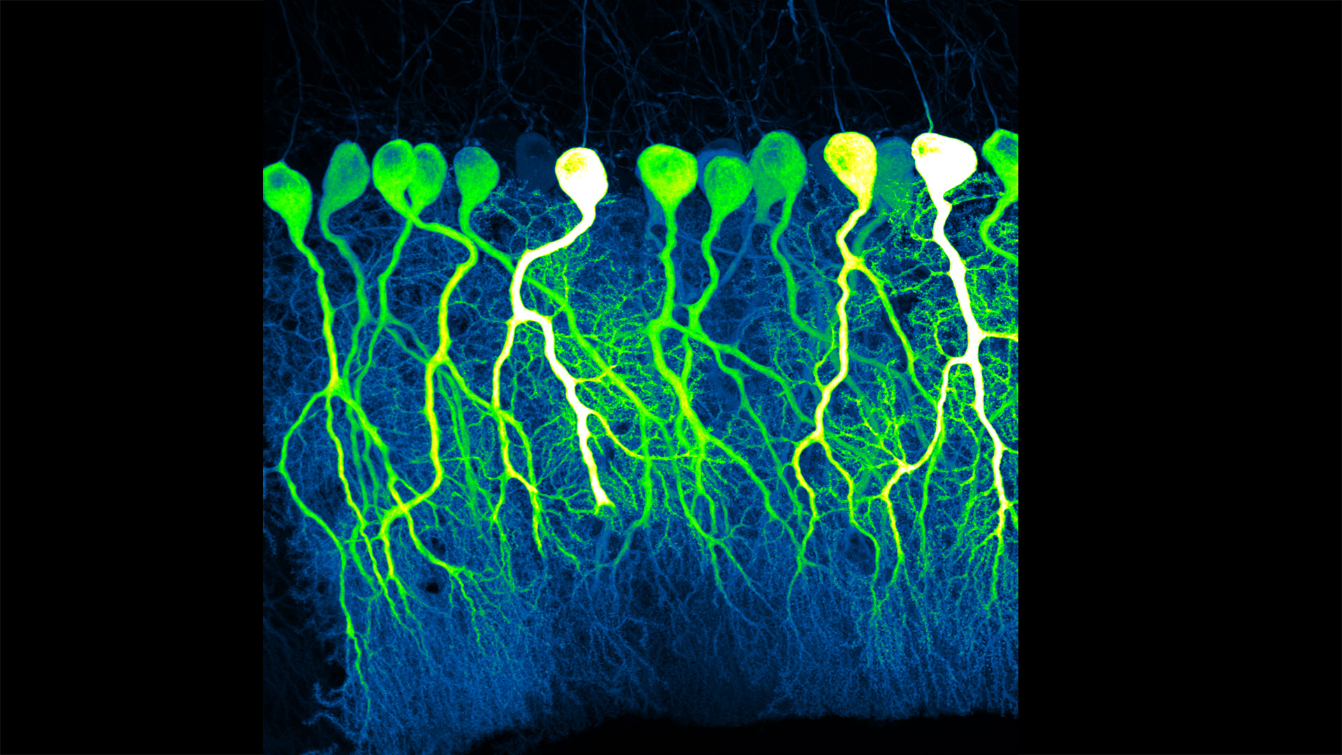
Silas Busch at the University of Chicago captured this slightly eerie scene, noting it reminded him of people shuffling through the dark of night. What you’re really seeing are some of the largest neurons in the mammalian brain, known as Purkinje cells. The photo won first place this year in the Brain Research through Advancing Innovative Neurotechnologies® (BRAIN) Initiative’s annual Show Us Your BRAINs! Photo and Video Contest.
While humans have them, too, the Purkinje cells pictured here are in the brain of a mouse. The head-like shapes you see in the image are the so-called soma, or the neurons’ cell bodies. Extending downwards are the heavily branched dendrites, which act like large antennae, receiving thousands of inputs from the rest of the body.
One reason this picture is such a standout, explains Busch, is because of what you don’t see. You’ll notice that only a few cells are fluorescently labeled and therefore lit up in green, leaving the rest in shadows. As a result, it’s possible to trace the detailed branches of individual Purkinje cells and make out their intricate forms. As it turns out, this ability to trace Purkinje cells so precisely led Busch, who is a graduate student in Christian Hansel’s lab focused on the neurobiology of learning and memory, to a surprising discovery, which the pair recently reported in Science.1
Purkinje cells connect to nerve fibers that “climb up” from the brain stem, which connects your brain and spinal cord to help control breathing, heart rate, balance and more. Scientists thought that each Purkinje cell received only one of these climbing fibers from the brain stem on its single primary branch.
However, after carefully tracing thousands of Purkinje cells in brain tissue from both mice and humans, the researchers have now shown that Purkinje cells and climbing fibers don’t always have a simple one-to-one relationship. In fact, Busch and Hansel found more than 95 percent of human Purkinje cells have multiple primary branches, not just one. In mice, that ratio is closer to 50 percent.
The researchers went on to show that mouse Purkinje cells with multiple primary branches often also receive multiple climbing fibers. The discovery rewrites the textbook idea of how Purkinje cells in the brain and climbing fibers from the brainstem are anatomically arranged.
Not surprisingly, those extra connections in the cerebellum (located in the back of the brain) also have important functional implications. When Purkinje cells have just one climbing fiber input, the new study shows, the whole cell receives each signal equally and responds accordingly. But, in cells with multiple climbing fiber inputs, the researchers could detect differences across a single cell depending on which primary branch received an input.
What this means is that Purkinje cells in the brain have much more computational power than had been appreciated. That extra brain power has important implications for understanding how brain circuits can adapt and respond to changes outside the body that now warrant further study. The new findings may have implications also for understanding the role of these Purkinje cell connections in some neurological and developmental disorders, including autism2 and a movement disorder known as cerebellar ataxia.
As they say, a picture is worth a thousand words. And this winning image comes as a reminder that we still have more to learn from careful study of basic brain anatomy, with important implications for human health and disease.
References:
[1] SE Busch and C Hansel. Climbing fiber multi-innervation of mouse Purkinje dendrites with arborization common to human. Science. DOI: 10.1126/science.adi1024. (2023).
[2] DH Simmons et al. Sensory Over-responsivity and Aberrant Plasticity in Cerebellar Cortex in a Mouse Model of Syndromic Autism. Biological Psychiatry: Global Open Science. DOI: 10.1016/j.bpsgos.2021.09.004. (2021).
Metabolomics: A New Approach to Understanding Glaucoma
Posted on by Michael F. Chiang, M.D., National Eye Institute

Glaucoma remains one of the most common causes of vision loss and blindness in the U.S. and much of the world, disproportionately affecting older people, African Americans, and Hispanics and Latinos. Early signs of glaucoma can vary, from eye pressure to changes in the appearance of the optic nerve, and the disease can progress for years undetected while causing irreversible vision loss. More research is needed to understand the complex processes that underpin how glaucoma develops and progresses. If detected early enough, doctors can intervene and stop or slow its progression, thus preventing or minimizing vision loss.
While more than 120 genetic factors have been linked to glaucoma, these genes account for less than 10% of glaucoma cases. Scientists are exploring other ways to predict glaucoma, including studying metabolites to see if they hold any clues. These small molecules are produced by metabolism, including the breakdown of nutrients when we digest food or byproducts from the medicine we take. Identifying at-risk individuals based on their metabolic profile might present an opportunity to intercept disease before vision loss.
Researchers already use metabolites as biomarkers or indicators to help diagnose disease or assess disease risk. There’s a standard blood test called a comprehensive metabolic blood panel that doctors use to measure levels of metabolites circulating in your blood—sugars like glucose, minerals such as calcium, and proteins such as creatinine.
Your metabolome is the complete set of metabolites not in just your blood but in your entire body. National Eye Institute-funded researchers led by Louis Pasquale, Icahn School of Medicine at Mount Sinai, New York, in collaboration with Oana A. Zeleznik and Jae Hee Kang of Brigham and Women’s Hospital, Boston, recently explored 369 blood metabolites in relation to glaucoma in a large study.1
The research team examined blood that had been stored frozen from two long-term studies of health professionals: the Nurses’ Health Studies and the Health Professionals’ Follow-Up Study. They compared about 600 participants who had developed glaucoma after study enrollment to a group of similar participants who didn’t. On average, the participants who developed glaucoma did so about 10 years after their initial blood draw in the study.
The researchers found a particularly strong association between glaucoma and two classes of lipids (fats): triglycerides and diglycerides. Patients with elevated triglycerides and diglycerides were more likely to develop glaucoma, and the association was strongest in a subtype of glaucoma that causes early loss of central vision. They confirmed their findings in a cross-sectional analysis of data from the UK Biobank.
High levels of triglycerides have been linked to a variety of health problems, notably heart disease and stroke. The good news is that effective treatments to control triglyceride levels already exist. Statin drugs, for example, lower blood lipid levels. While studies looking at statin use and glaucoma risk have shown mixed results, we may learn that specific subtypes of glaucoma can be effectively controlled with statins. More research is needed to know if existing drugs might prevent glaucoma.
Pasquale’s work adds to a growing body of evidence that links health status to metabolism. Similar associations have been made between various metabolites and kidney cancer,2 pregnancy complications,3 type 2 diabetes,4 and Alzheimer’s disease.5 For researchers interested in exploring associations between metabolites and disease risk, the NIH Common Fund offers scientists a national and international repository for metabolomics data and metadata called the Metabolomics Workbench Metabolite Database, which contained more than 167,000 entries in 2022.
These findings and others offer the potential to prevent more and treat less. We urge anyone in an at-risk group, including people with a family history of glaucoma, to get regular, comprehensive eye exams.
References:
[1] OA Zeleznik, et al. Plasma metabolite profile for primary open-angle glaucoma in three US cohorts and the UK Biobank. Nature Communications DOI:10.1038/s41467-023-38466-x (2023)
[2] OO Bifarin, et al. Urine-Based Metabolomics and Machine Learning Reveals Metabolites Associated with Renal Cell Carcinoma Stage. Cancers (Basel) DOI:10.3390/cancers13246253 (2021)
[3] EW Harville, et al. Untargeted analysis of first trimester serum to reveal biomarkers of pregnancy complications: a case-control discovery phase study. Scientific Reports DOI:10.1038/s41598-021-82804-1 (2021)
[4] Nightingale Health Biobank Collaborative Group, et al. Metabolomic and genomic prediction of common diseases in 477,706 participants in three national biobanks. medRxiv DOI: 10.1101/2023.06.09.23291213 (2023). *note this article is a pre-print and is not peer-reviewed
[5] DK Barupal, et al. Sets of coregulated serum lipids are associated with Alzheimer’s disease pathophysiology. Alzheimer’s & Dementia: Diagnosis, Assessment & Disease Monitoring. DOI:10.1016/j.dadm.2019.07.002 (2019)
NIH Support: National Eye Institute, National Cancer Institute
Editor’s note: This blog post was updated on Jan. 18, 2024, to include Oana A. Zeleznik as one of the collaborators.
Reflecting on Two Years of Discovery and Looking Ahead to New NIH Leadership
Posted on by Lawrence Tabak, D.D.S., Ph.D.

As I transition from my role as the Acting NIH Director, I’d like to thank you, the readers, for visiting the NIH Director’s Blog ever since I took the helm 22 months ago. From Long COVID to the opioid overdose epidemic to Alzheimer’s disease—we’ve covered a range of diseases and conditions, scientific advances, and programs. You were able to read about such a broad spectrum of science thanks in large part to the many Institute Directors at NIH who authored guest posts. I hope the blog has helped you learn more about what NIH does and the many ways that biomedical research impacts human health.
A key focus of my career as both a scientific investigator and administrative leader has been supporting trainees and finding new ways to cultivate and expand the next generation of researchers. In my many discussions with young investigators, I’ve often reminded them that they should not be afraid to fail. To the students and early-stage scientists who have visited this site: I hope these stories of discovery—often the result of earlier failures—have provided some insight and inspiration as you move through your scientific career or consider starting one.
I’d also like to thank the many people—employees, government and private partners, patients, scientists, advocates, and other members of the public—who have reached out with messages of support, and sometimes with messages of criticism. Both have helped inform the decisions I needed to make to fulfill the NIH mission.
In closing, I congratulate Dr. Monica Bertagnolli as she takes the helm as the next permanent NIH director. Dr. Bertagnolli—an outstanding physician scientist—is a strong leader who will bring fresh, bold new ideas to NIH and the biomedical research enterprise. I know she’ll be in good hands thanks to the outstanding staff across NIH and the leadership in the Department of Health and Human Services. I look forward to supporting her efforts and continuing to ensure that NIH research optimizes health for all people.
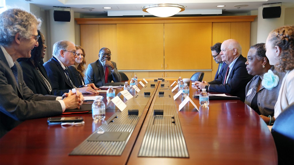
Senator Ben Cardin Visits NIH
Posted on by Lawrence Tabak, D.D.S., Ph.D.
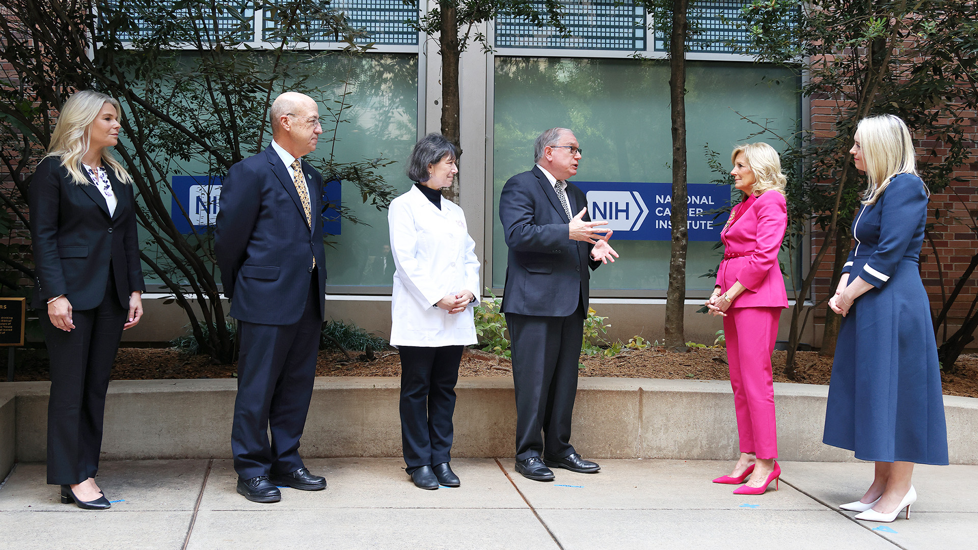
First Lady Dr. Jill Biden Visits NIH
Posted on by Lawrence Tabak, D.D.S., Ph.D.
How Double-Stranded RNA Protects the Brain Against Infection While Making Damaging Neuroinflammation More Likely
Posted on by Lawrence Tabak, D.D.S., Ph.D.

When you get a run-of-the-mill viral infection, after a few days of symptoms your immune system typically fends off the bug, and you’ll make a full recovery. In rare cases, a virus can infect the brain. This can lead to much bigger problems, including cognitive impairments known as “brain fog,” other neuropsychiatric symptoms, potentially irreversible brain damage, or even death. For this reason, the brain, more than other parts of the body, relies heavily on immune responses that can control viral infections immediately.
Now some intriguing findings from an NIH-funded team reported in Science Immunology help to explain how the brain is protected against infections.1 However, the findings also highlight a serious downside: these same mechanisms that protect the brain also leave it especially vulnerable to damaging levels of neuroinflammation.
The new findings may help to explain what goes on in the brains of people with a wide range of neurodegenerative conditions, including amyotrophic lateral sclerosis (ALS) and Alzheimer’s disease. They also point to promising targets for developing treatments that might turn inflammatory immune responses in the brain up or down, as desired, to treat these and other serious conditions.
How does it work? The key is double-stranded RNA (dsRNA).
RNA molecules are readouts of genetic information in DNA that carry instructions for building the proteins that carry out various cell functions. RNA molecules in our cells are most often in single-stranded or short dsRNA form. In contrast, lengthy dsRNAs are a hallmark of viruses. When a virus invades our cells, our immune system’s first line of defense can sense those long viral dsRNAs and trigger a response.
But it turns out that dsRNAs aren’t unique to viruses, as the new study highlights. The researchers, led by Tyler Dorrity and Heegwon Shin, both members of Hachung Chung’s lab at Columbia University Irving Medical Center, New York, found that human neurons—even when they’re normal and healthy—also have exceptionally high levels of long dsRNAs.
Their lab studies in cells and tissues show that these dsRNAs in neurons can trigger an inflammatory immune response just as they do in viruses. By manipulating neurons in a way that cut back on the number of dsRNAs, they found they could lower the innate immune response. However, cells with fewer dsRNAs also showed greater susceptibility to infection with Zika viruses and herpes simplex virus, which can produce a form of viral encephalitis.
The researchers also knew from earlier studies that people with a rare, inherited condition called Aicardi-Goutières syndrome (AGS), which primarily affects the brain and immune system, carry a mutation that causes their cells to lack an enzyme needed to edit dsRNAs. As a result, neurons carrying this mutation have so many dsRNAs that it is toxic.
They went on to show that they could shift this dynamic by altering levels of two other proteins that bind RNA. The proteins normally encourage dsRNA formation in the brain. When the researchers deleted these RNA-binding proteins from the AGS neurons, those neurons made fewer long dsRNAs, which in turn protected them from the inflammatory immune responses and allowed them to survive longer. As expected, however, those cells also were more susceptible to viral infection.
The findings show how this tricky balance between susceptibility to infection and inflammation in the brain works in both health and disease. It also leads to the tantalizing suggestion that treatments targeting these various players or others in the same pathways may offer new ways of treating brain infections or neuroinflammatory conditions, by boosting or dampening dsRNA levels and the associated immune responses. As a next step, the researchers report that they’re pursuing studies to explore the role of dsRNA-triggered immune responses in ALS and Alzheimer’s, as well as in neuropsychiatric symptoms sometimes seen in people with lupus.
References:
[1] TJ Dorrity TJ, et al. Long 3’UTRs predispose neurons to inflammation by promoting immunostimulatory double-stranded RNA formation. Science Immunology DOI: 10.1126/sciimmunol.adg2979 (2023).
NIH Support: National Institute of Neurological Disorders and Stroke, National Institute of Allergy and Infectious Diseases, National Institute of General Medical Sciences
Brain Atlas Paves the Way for New Understanding of How the Brain Functions
Posted on by Lawrence Tabak, D.D.S., Ph.D.

When NIH launched The BRAIN Initiative® a decade ago, one of many ambitious goals was to develop innovative technologies for profiling single cells to create an open-access reference atlas cataloguing the human brain’s many parts. The ultimate goal wasn’t to produce a single, static reference map, but rather to capture a dynamic view of how the brain’s many cells of varied types are wired to work together in the healthy brain and how this picture may shift in those with neurological and mental health disorders.
So I’m now thrilled to report the publication of an impressive collection of work from hundreds of scientists in the BRAIN Initiative Cell Census Network (BICCN), detailed in more than 20 papers in Science, Science Advances, and Science Translational Medicine.1 Among many revelations, this unprecedented, international effort has characterized more than 3,000 human brain cell types. To put this into some perspective, consider that the human lung contains 61 cell types.2 The work has also begun to uncover normal variation in the brains of individual people, some of the features that distinguish various disease states, and distinctions among key parts of the human brain and those of our closely related primate cousins.
Of course, it’s not possible to do justice to this remarkable body of work or its many implications in the space of a single blog post. But to give you an idea of what’s been accomplished, some of these studies detail the primary effort to produce a comprehensive brain atlas, including defining the brain’s many cell types along with their underlying gene activity and the chemical modifications that turn gene activity up or down.3,4,5
Other studies in this collection take a deep dive into more specific brain areas. For instance, to capture normal variations among people, a team including Nelson Johansen, University of California, Davis, profiled cells in the neocortex—the outermost portion of the brain that’s responsible for many complex human behaviors.6 Overall, the work revealed a highly consistent cellular makeup from one person to the next. But it also highlighted considerable variation in gene activity, some of which could be explained by differences in age, sex and health. However, much of the observed variation remains unexplained, opening the door to more investigations to understand the meaning behind such brain differences and their role in making each of us who we are.
Yang Li, now at Washington University in St. Louis, and his colleagues analyzed 1.1 million cells from 42 distinct brain areas in samples from three adults.4 They explored various cell types with potentially important roles in neuropsychiatric disorders and were able to pinpoint specific cell types, genes and genetic switches that may contribute to the development of certain traits and disorders, including bipolar disorder, depression and schizophrenia.
Yet another report by Nikolas Jorstad, Allen Institute, Seattle, and colleagues delves into essential questions about what makes us human as compared to other primates like chimpanzees.7 Their comparisons of gene activity at the single-cell level in a specific area of the brain show that humans and other primates have largely the same brain cell types, but genes are activated differently in specific cell types in humans as compared to other primates. Those differentially expressed genes in humans often were found in portions of the genome that show evidence of rapid change over evolutionary time, suggesting that they play important roles in human brain function in ways that have yet to be fully explained.
All the data represented in this work has been made publicly accessible online for further study. Meanwhile, the effort to build a more finely detailed picture of even more brain cell types and, with it, a more complete understanding of human brain circuitry and how it can go awry continues in the BRAIN Initiative Cell Atlas Network (BICAN). As impressive as this latest installment is—in our quest to understand the human brain, brain disorders, and their treatment—we have much to look forward to in the years ahead.
References:
A list of all the papers part of the brain atlas research is available here: https://www.science.org/collections/brain-cell-census.
[1] M Maroso. A quest into the human brain. Science DOI: 10.1126/science.adl0913 (2023).
[2] L Sikkema, et al. An integrated cell atlas of the lung in health and disease. Nature Medicine DOI: 10.1038/s41591-023-02327-2 (2023).
[3] K Siletti, et al. Transcriptomic diversity of cell types across the adult human brain. Science DOI: 10.1126/science.add7046 (2023).
[4] Y Li, et al. A comparative atlas of single-cell chromatin accessibility in the human brain. Science DOI: 10.1126/science.adf7044 (2023).
[5] W Tian, et al. Single-cell DNA methylation and 3D genome architecture in the human brain. Science DOI: 10.1126/science.adf5357 (2023).
[6] N Johansen, et al. Interindividual variation in human cortical cell type abundance and expression. Science DOI: 10.1126/science.adf2359 (2023).
[7] NL Jorstad, et al. Comparative transcriptomics reveals human-specific cortical features. Science DOI: 10.1126/science.ade9516 (2023).
NIH Support: Projects funded through the NIH BRAIN Initiative Cell Consensus Network
Can Bioprinted Skin Substitutes Replace Traditional Grafts for Treating Burn Injuries and Other Serious Skin Wounds?
Posted on by Lawrence Tabak, D.D.S., Ph.D.
Each year in the U.S., more than 500,000 people receive treatment for burn injuries and other serious skin wounds.1 To close the most severe wounds with less scarring, doctors often must surgically remove skin from one part of a person’s body and use it to patch the injured site. However, this is an intensive process, and some burn patients with extensive skin loss do not have sufficient skin available for grafting. Scientists have been exploring ways to repair these serious skin wounds without skin graft surgery.
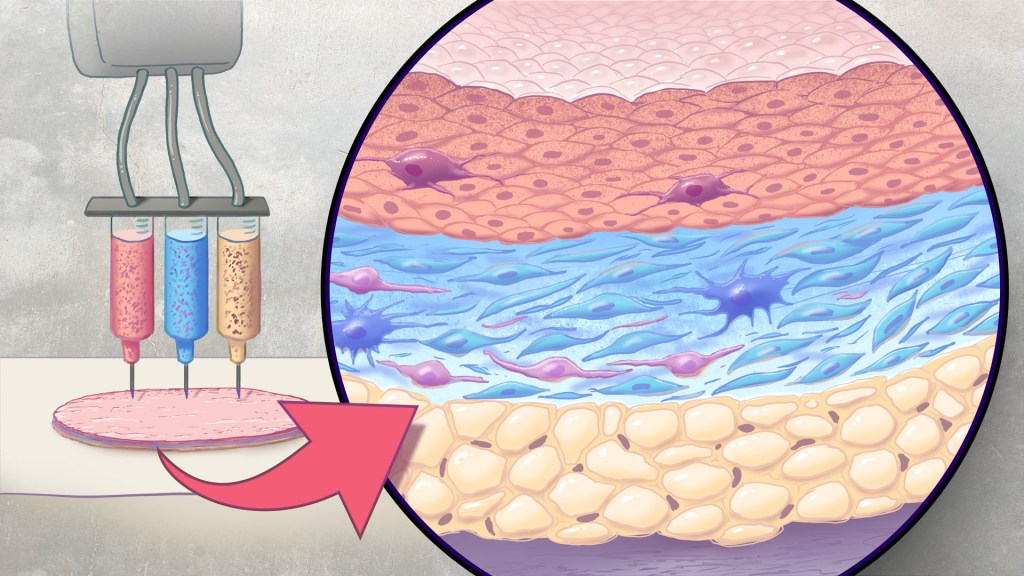
An NIH-funded team recently showed that bioprinted skin substitutes may serve as a promising alternative to traditional skin grafts in preclinical studies reported in Science Translational Medicine.2 The approach involves a portable skin bioprinter system that deposits multiple layers of skin directly into a wound. The recent findings add to evidence that bioprinting technology can successfully regenerate human-like skin to allow healing. While this approach has yet to be tested in people, it confirms that such technologies already can produce skin constructs with the complex structures and multiple cell types present in healthy human skin.
This latest work comes from a team led by Adam Jorgensen and Anthony Atala at Wake Forest School of Medicine’s Wake Forest Institute for Regenerative Medicine, Winston-Salem, NC. Members of the Atala lab and their colleagues had earlier shown it was possible to isolate two major skin cell types found in the skin’s outer (epidermis) and middle (dermis) layers from a small biopsy of healthy skin, expand the number of cells in the lab and then deliver the cells directly into an injury using a specially designed bioprinter.3 Using integrated imaging technology to scan a wound, computer software “prints” cells right into an injury, mimicking two of our skin’s three natural layers.
In the new study, Atala’s team has gone even further to construct skin substitutes that mimic the structure of human skin and that include six primary human skin cell types. They then used their bioprinter to produce skin constructs with all three layers found in healthy human skin: epidermis, dermis, and hypodermis.
To put their skin substitutes to the test, they first transplanted them into mice. Their studies showed that the bioprinted skin encouraged the rapid growth of new blood vessels and had other features of normal-looking, healthy skin. The researchers were able to confirm that their bioprinted skin implants successfully integrated into the animals’ regenerated skin to speed healing.
Studies in a pig model of wound healing added to evidence that such bioprinted implants can successfully repair full-thickness wounds, meaning those that extend through all three layers of skin. The bioprinted skin patches allowed for improved wound healing with less scarring. They also found that the bioprinted grafts encouraged activity in the skin from genes known to play important roles in wound healing.
It’s not yet clear if this approach will work as well in the clinic as it does in the lab. To make it feasible, the researchers note there’s a need for improved approaches to isolating and expanding the needed skin cell types. Nevertheless, these advances come as encouraging evidence that bioprinted skin substitutes could one day offer a promising alternative to traditional skin grafts with the capacity to help even more people with severe burns or other wounds.
References:
[1] Burn Incidence Fact Sheet. American Burn Association
[2] AM Jorgensen, et al. Multicellular bioprinted skin facilitates human-like skin architecture in vivo. Science Translational Medicine DOI: 10.1126/scitranslmed.adf7547 (2023).
[3] M Albanna, et al. In Situ Bioprinting of Autologous Skin Cells Accelerates Wound Healing of Extensive Excisional Full-Thickness Wounds. Scientific Reports DOI: 10.1038/s41598-018-38366-w (2019).
NIH Support: National Institute of Arthritis and Musculoskeletal and Skin Diseases
Previous Page Next Page