196 Search Results for "covid-19"
U.K. Study Shows Power of Digital Contact Tracing for COVID-19
Posted on by Dr. Francis Collins

There’s been much interest in using digital technology to help contain the spread of COVID-19 in our communities. The idea is to make available opt-in smart phone apps that create a log of other apps operating on the phones of nearby participants. If a participant tests positive for COVID-19 and enters the result, the app will then send automatic alerts to those phones—and participants—who recently came into close proximity with them.
In theory, digital tracing would be much faster and more efficient than the challenging detective work involved in traditional contract tracing. But many have wondered how well such an opt-in system would work in practice. A recent paper, published in the journal Nature, shows that a COVID-19 digital tracing app worked quite well in the United Kingdom [1].
The research comes from Christophe Fraser, Oxford University, and his colleagues in the U.K. The team studied the NHS COVID-19 app, the National Health Service’s digital tracing smart phone app for England and Wales. Launched in September 2020, the app has been downloaded onto 21 million devices and used regularly by about half of eligible smart phone users, ages 16 and older. That’s 16.5 million of 33.7 million people, or more than a quarter of the total population of England and Wales.
From the end of September through December 2020, the app sent about 1.7 million exposure notifications. That’s 4.4 on average for every person with COVID-19 who opted-in to the digital tracing app.
The researchers estimate that around 6 percent of app users who received notifications of close contact with a positive case went on to test positive themselves. That’s similar to what’s been observed in traditional contact tracing.
Next, they used two different approaches to construct mathematical and statistical models to determine how likely it was that a notified contact, if infected, would quarantine in a timely manner. Though the two approaches arrived at somewhat different answers, their combined outputs suggest that the app may have stopped anywhere from 200,000 to 900,000 infections in just three months. This means that roughly one case was averted for each COVID-19 case that consented to having their contacts notified through the app.
Of course, these apps are only as good as the total number of people who download and use them faithfully. They estimate that for every 1 percent increase in app users, the number of COVID-19 cases could be reduced by another 1 or 2 percent. While those numbers might sound small, they can be quite significant when one considers the devastating impact that COVID-19 continues to have on the lives and livelihoods of people all around the world.
Reference:
[1] The epidemiological impact of the NHS COVID-19 App. Wymant C, Ferretti L, Tsallis D, Charalambides M, Abeler-Dörner L, Bonsall D, Hinch R, Kendall M, Milsom L, Ayres M, Holmes C, Briers M, Fraser C. Nature. 2021 May 12.
Links:
COVID-19 Research (NIH)
Christophe Fraser (Oxford University, UK)
Giving Thanks to Everyone at NIH’s COVID-19 Vaccine Clinic
Posted on by Dr. Francis Collins

How Severe COVID-19 Can Tragically Lead to Lung Failure and Death
Posted on by Dr. Francis Collins
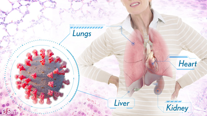
More than 3 million people around the world, now tragically including thousands every day in India, have lost their lives to severe COVID-19. Though incredible progress has been made in a little more than a year to develop effective vaccines, diagnostic tests, and treatments, there’s still much we don’t know about what precisely happens in the lungs and other parts of the body that leads to lethal outcomes.
Two recent studies in the journal Nature provide some of the most-detailed analyses yet about the effects on the human body of SARS-CoV-2, the coronavirus that causes COVID-19 [1,2]. The research shows that in people with advanced infections, SARS-CoV-2 often unleashes a devastating series of host events in the lungs prior to death. These events include runaway inflammation and rampant tissue destruction that the lungs cannot repair.
Both studies were supported by NIH. One comes from a team led by Benjamin Izar, Columbia University, New York. The other involves a group led by Aviv Regev, now at Genentech, and formerly at Broad Institute of MIT and Harvard, Cambridge, MA.
Each team analyzed samples of essential tissues gathered from COVID-19 patients shortly after their deaths. Izar’s team set up a rapid autopsy program to collect and freeze samples within hours of death. He and his team performed single-cell RNA sequencing on about 116,000 cells from the lung tissue of 19 men and women. Similarly, Regev’s team developed an autopsy biobank that included 420 total samples from 11 organ systems, which were used to generate multiple single-cell atlases of tissues from the lung, kidney, liver, and heart.
Izar’s team found that the lungs of people who died of COVID-19 were filled with immune cells called macrophages. While macrophages normally help to fight an infectious virus, they seemed in this case to produce a vicious cycle of severe inflammation that further damaged lung tissue. The researchers also discovered that the macrophages produced high levels of IL-1β, a type of small inflammatory protein called a cytokine. This suggests that drugs to reduce effects of IL-1β might have promise to control lung inflammation in the sickest patients.
As a person clears and recovers from a typical respiratory infection, such as the flu, the lung repairs the damage. But in severe COVID-19, both studies suggest this isn’t always possible. Not only does SARS-CoV-2 destroy cells within air sacs, called alveoli, that are essential for the exchange of oxygen and carbon dioxide, but the unchecked inflammation apparently also impairs remaining cells from repairing the damage. In fact, the lungs’ regenerative cells are suspended in a kind of reparative limbo, unable to complete the last steps needed to replace healthy alveolar tissue.
In both studies, the lung tissue also contained an unusually large number of fibroblast cells. Izar’s team went a step further to show increased numbers of a specific type of pathological fibroblast, which likely drives the rapid lung scarring (pulmonary fibrosis) seen in severe COVID-19. The findings point to specific fibroblast proteins that may serve as drug targets to block deleterious effects.
Regev’s team also describes how the virus affects other parts of the body. One surprising discovery was there was scant evidence of direct SARS-CoV-2 infection in the liver, kidney, or heart tissue of the deceased. Yet, a closer look heart tissue revealed widespread damage, documenting that many different coronary cell types had altered their genetic programs. It’s still to be determined if that’s because the virus had already been cleared from the heart prior to death. Alternatively, the heart damage might not be caused directly by SARS-CoV-2, and may arise from secondary immune and/or metabolic disruptions.
Together, these two studies provide clearer pictures of the pathology in the most severe and lethal cases of COVID-19. The data from these cell atlases has been made freely available for other researchers around the world to explore and analyze. The hope is that these vast data sets, together with future analyses and studies of people who’ve tragically lost their lives to this pandemic, will improve our understanding of long-term complications in patients who’ve survived. They also will now serve as an important foundational resource for the development of promising therapies, with the goal of preventing future complications and deaths due to COVID-19.
References:
[1] A molecular single-cell lung atlas of lethal COVID-19. Melms JC, Biermann J, Huang H, Wang Y, Nair A, Tagore S, Katsyv I, Rendeiro AF, Amin AD, Schapiro D, Frangieh CJ, Luoma AM, Filliol A, Fang Y, Ravichandran H, Clausi MG, Alba GA, Rogava M, Chen SW, Ho P, Montoro DT, Kornberg AE, Han AS, Bakhoum MF, Anandasabapathy N, Suárez-Fariñas M, Bakhoum SF, Bram Y, Borczuk A, Guo XV, Lefkowitch JH, Marboe C, Lagana SM, Del Portillo A, Zorn E, Markowitz GS, Schwabe RF, Schwartz RE, Elemento O, Saqi A, Hibshoosh H, Que J, Izar B. Nature. 2021 Apr 29.
[2] COVID-19 tissue atlases reveal SARS-CoV-2 pathology and cellular targets. Delorey TM, Ziegler CGK, Heimberg G, Normand R, Shalek AK, Villani AC, Rozenblatt-Rosen O, Regev A. et al. Nature. 2021 Apr 29.
Links:
COVID-19 Research (NIH)
Izar Lab (Columbia University, New York)
Aviv Regev (Genentech, South San Francisco, CA)
NIH Support: National Center for Advancing Translational Sciences; National Heart, Lung, and Blood Institute; National Cancer Institute; National Institute of Allergy and Infectious Diseases; National Institute of Diabetes and Digestive and Kidney Diseases; National Human Genome Research Institute; National Institute of Mental Health; National Institute on Alcohol Abuse and Alcoholism
Dynamic View of Spike Protein Reveals Prime Targets for COVID-19 Treatments
Posted on by Dr. Francis Collins

This striking portrait features the spike protein that crowns SARS-CoV-2, the coronavirus that causes COVID-19. This highly flexible protein has settled here into one of its many possible conformations during the process of docking onto a human cell before infecting it.
This portrait, however, isn’t painted on canvas. It was created on a computer screen from sophisticated 3D simulations of the spike protein in action. The aim was to map its many shape-shifting maneuvers accurately at the atomic level in hopes of detecting exploitable structural vulnerabilities to thwart the virus.
For example, notice the many chain-like structures (green) that adorn the protein’s surface (white). They are sugar molecules called glycans that are thought to shield the spike protein by sweeping away antibodies. Also notice areas (purple) that the simulation identified as the most-attractive targets for antibodies, based on their apparent lack of protection by those glycans.
This work, published recently in the journal PLoS Computational Biology [1], was performed by a German research team that included Mateusz Sikora, Max Planck Institute of Biophysics, Frankfurt. The researchers used a computer application called molecular dynamics (MD) simulation to power up and model the conformational changes in the spike protein on a time scale of a few microseconds. (A microsecond is 0.000001 second.)
The new simulations suggest that glycans act as a dynamic shield on the spike protein. They liken them to windshield wipers on a car. Rather than being fixed in space, those glycans sweep back and forth to protect more of the protein surface than initially meets the eye.
But just as wipers miss spots on a windshield that lie beyond their tips, glycans also miss spots of the protein just beyond their reach. It’s those spots that the researchers suggest might be prime targets on the spike protein that are especially promising for the design of future vaccines and therapeutic antibodies.
This same approach can now be applied to identifying weak spots in the coronavirus’s armor. It also may help researchers understand more fully the implications of newly emerging SARS-CoV-2 variants. The hope is that by capturing this devastating virus and its most critical proteins in action, we can continue to develop and improve upon vaccines and therapeutics.
Reference:
[1] Computational epitope map of SARS-CoV-2 spike protein. Sikora M, von Bülow S, Blanc FEC, Gecht M, Covino R, Hummer G. PLoS Comput Biol. 2021 Apr 1;17(4):e1008790.
Links:
COVID-19 Research (NIH)
Mateusz Sikora (Max Planck Institute of Biophysics, Frankfurt, Germany)
The surprising properties of the coronavirus envelope (Interview with Mateusz Sikora), Scilog, November 16, 2020.
A Real-World Look at COVID-19 Vaccines Versus New Variants
Posted on by Dr. Francis Collins

Clinical trials have shown the COVID-19 vaccines now being administered around the country are highly effective in protecting fully vaccinated individuals from the coronavirus SARS-CoV-2. But will they continue to offer sufficient protection as the frequency of more transmissible and, in some cases, deadly emerging variants rise?
More study and time is needed to fully answer this question. But new data from Israel offers an early look at how the Pfizer/BioNTech vaccine is holding up in the real world against coronavirus “variants of concern,” including the B.1.1.7 “U.K. variant” and the B.1.351 “South African variant.” And, while there is some evidence of breakthrough infections, the findings overall are encouraging.
Israel was an obvious place to look for answers to breakthrough infections. By last March, more than 80 percent of the country’s vaccine-eligible population had received at least one dose of the Pfizer/BioNTech vaccine. An earlier study in Israel showed that the vaccine offered 94 percent to 96 percent protection against infection across age groups, comparable to the results of clinical trials. But it didn’t dig into any important differences in infection rates with newly emerging variants, post-vaccination.
To dig a little deeper into this possibility, a team led by Adi Stern, Tel Aviv University, and Shay Ben-Shachar, Clalit Research Institute, Tel Aviv, looked for evidence of breakthrough infections in several hundred people who’d had at least one dose of the Pfizer/BioNTech vaccine [1]. The idea was, if this vaccine were less effective in protecting against new variants of concern, the proportion of infections caused by them should be higher in vaccinated compared to unvaccinated individuals.
During the study, reported as a pre-print in MedRxiv, it became clear that B.1.1.7 was the predominant SARS-CoV-2 variant in Israel, with its frequency increasing over time. By comparison, the B.1.351 “South African” variant was rare, accounting for less than 1 percent of cases sampled in the study. No other variants of concern, as defined by the World Health Organization, were detected.

In total, the researchers sequenced SARS-CoV-2 from more than 800 samples, including vaccinated individuals and matched unvaccinated individuals with similar characteristics including age, sex, and geographic location. They identified nearly 250 instances in which an individual became infected with SARS-CoV-2 after receiving their first vaccine dose, meaning that they were only partially protected. Almost 150 got infected sometime after receiving the second dose.
Interestingly, the evidence showed that these breakthrough infections with the B.1.1.7 variant occurred slightly more often in people after the first vaccine dose compared to unvaccinated people. No evidence was found for increased breakthrough rates of B.1.1.7 a week or more after the second dose. In contrast, after the second vaccine dose, infection with the B.1.351 became slightly more frequent. The findings show that people remain susceptible to B.1.1.7 following a single dose of vaccine. They also suggest that the two-dose vaccine may be slightly less effective against B.1.351 compared to the original or B.1.1.7 variants.
It’s important to note, however, that the researchers only observed 11 infections with the B.1.351 variant—eight of them in individuals vaccinated with two doses. Interestingly, all eight tested positive seven to 13 days after receiving their second dose. No one in the study tested positive for this variant two weeks or more after the second dose.
Many questions remain, including whether the vaccines reduced the duration and/or severity of infections. Nevertheless, the findings are a reminder that—while these vaccines offer remarkable protection—they are not foolproof. Breakthrough infections can and do occur.
In fact, in a recent report in the New England Journal of Medicine, NIH-supported researchers detailed the experiences of two fully vaccinated individuals in New York who tested positive for COVID-19 [2]. Though both recovered quickly at home, genomic data in those cases revealed multiple mutations in both viral samples, including a variant first identified in South Africa and Brazil, and another, which has been spreading in New York since November.
These findings in Israel and the United States also highlight the importance of tracking coronavirus variants and making sure that all eligible individuals get fully vaccinated as soon as they have the opportunity. They show that COVID-19 testing will continue to play an important role, even in those who’ve already been vaccinated. This is even more important now as new variants continue to rise in frequency.
Just over 100 million Americans aged 18 and older—about 40 percent of adults—are now fully vaccinated [3]. However, we need to get that number much higher. If you or a loved one haven’t yet been vaccinated, please consider doing so. It will help to save lives and bring this pandemic to an end.
References:
[1] Evidence for increased breakthrough rates of SARS-CoV-2 variants of concern in BNT162b2 mRNA vaccinated individuals. Kustin T et al. medRxiv. April 16, 2021.
[2] Vaccine breakthrough infections with SARS-CoV-2 variants. Hacisuleyman E, Hale C, Saito Y, Blachere NE, Bergh M, Conlon EG, Schaefer-Babajew DJ, DaSilva J, Muecksch F, Gaebler C, Lifton R, Nussenzweig MC, Hatziioannou T, Bieniasz PD, Darnell RB. N Engl J Med. 2021 Apr 21.
[3] COVID-19 vaccinations in the United States. Centers for Disease Control and Prevention.
Links:
COVID-19 Research (NIH)
Stern Lab (Tel Aviv University, Israel)
Ben-Shachar Lab (Clalit Research Institute, Tel Aviv, Israel)
NIH Support: National Institute of Allergy and Infectious Diseases
Building Confidence in COVID-19 Vaccines
Posted on by Dr. Francis Collins

Learning from History: Fauci Donates Model to Smithsonian’s COVID-19 Collection
Posted on by Dr. Francis Collins
Not too long after the global coronavirus disease 2019 (COVID-19) pandemic reached the United States, museum curators began collecting material to document the history of this devastating public health crisis and our nation’s response to it. To help tell this story, the Smithsonian Institution’s National Museum of American History recently scored a donation from my friend and colleague Dr. Anthony Fauci, Director of NIH’s National Institute of Allergy and Infectious Diseases.
Widely recognized for serving as a clear voice for science throughout the pandemic, Fauci gave the museum his much-used model of SARS-CoV-2, which is the coronavirus that causes COVID-19. This model, which is based on work conducted by NIH-supported electron microscopists and structural biologists, was 3D printed right here at NIH. By the way, I’m lucky enough to have one too.
Both of these models have “met” an amazing array of people—from presidents to congresspeople to journalists to average citizens—as part of our efforts to help folks understand SARS-CoV-2 and the crucial role of its surface spike proteins. As shown in this brief video, Fauci raised his model one last time and then, ever the public ambassador for science, turned his virtual donation into a memorable teaching moment. I recommend you take a minute or two to watch it.
The donation took place during a virtual ceremony in which the National Museum of American History awarded Fauci its prestigious Great Americans Medal. He received the award for his lifetime contributions to the nation’s ideals and for making a lasting impact on public health via his many philanthropic and humanitarian efforts. Fauci joined an impressive list of luminaries in receiving this honor, including former Secretaries of State Madeleine Albright and General Colin Powell; journalist Tom Brokaw; baseball great Cal Ripken Jr.; tennis star Billie Jean King; and musician Paul Simon. It’s a well-deserved honor for a physician-scientist who’s advised seven presidents on a range of domestic and global health issues, from HIV/AIDS to Ebola to COVID-19.
With Fauci’s model now enshrined as an official piece of U.S. history, the Smithsonian and other museums around the world are stepping up their efforts to gather additional artifacts related to COVID-19 and to chronicle its impacts on the health and economy of our nation. Hopefully, future generations will learn from this history so that humankind is not doomed to repeat it.
It is interesting to note that the National Museum of American History’s collection contains few artifacts from another tragic chapter in our nation’s past: the 1918 Influenza Pandemic. One reason this pandemic went largely undocumented is that, like so many of their fellow citizens, curators chose to overlook its devastating impacts and instead turn toward the future.

Today, museum staffers across the country and around the world are stepping up to the challenge of documenting COVID-19’s history with great creativity, collecting all variety of masks, test kits, vaccine vials, and even a few ventilators. At the NIH’s main campus in Bethesda, MD, the Office of NIH History and Stetten Museum is busy preparing a small exhibit of scientific and clinical artifacts that could open as early as the summer of 2021. The museum is also collecting oral histories as part of its “Behind the Mask” project. So far, more than 50 interviews have been conducted with NIH staff, including a scientist who’s helping the hard-hit Navajo Nation during the pandemic; a Clinical Center nurse who’s treating patients with COVID-19, and a mental health professional who’s had to change expectations since the outbreak.
The pandemic isn’t over yet. All of us need to do our part by getting vaccinated against COVID-19 and taking other precautions to prevent the virus’s deadly spread. But won’t it great when—hopefully, one day soon—we can relegate this terrible pandemic to the museums and the history books!
Links:
COVID-19 Research (NIH)
Video: National Museum of American History Presents The Great Americans Medal to Anthony S. Fauci (Smithsonian Institution, Washington, D.C.)
National Museum of American History (Smithsonian)
The Office of NIH History and Stetten Museum (NIH)
Mapping Severe COVID-19 in the Lungs at Single-Cell Resolution
Posted on by Dr. Francis Collins

A crucial question for COVID-19 researchers is what causes progression of the initial infection, leading to life-threatening respiratory illness. A good place to look for clues is in the lungs of those COVID-19 patients who’ve tragically lost their lives to acute respiratory distress syndrome (ARDS), in which fluid and cellular infiltrates build up in the lung’s air sacs, called alveoli, keeping them from exchanging oxygen with the bloodstream.
As shown above, a team of NIH-funded researchers has done just that, capturing changes in the lungs over the course of a COVID-19 infection at unprecedented, single-cell resolution. These imaging data add evidence that SARS-CoV-2, the coronavirus that causes COVID-19, primarily infects cells at the surface of the air sacs. Their findings also offer valuable clues for treating the most severe consequences of COVID-19, suggesting that a certain type of scavenging immune cell might be driving the widespread lung inflammation that leads to ARDS.
The findings, published in Nature [1], come from Olivier Elemento and Robert E. Schwartz, Weill Cornell Medicine, New York. They already knew from earlier COVID-19 studies about the body’s own immune response causing the lung inflammation that leads to ARDS. What was missing was an understanding of the precise interplay between immune cells and lung tissue infected with SARS-CoV-2. It also wasn’t clear how the ARDS seen with COVID-19 compared to the ARDS seen in other serious respiratory diseases, including influenza and bacterial pneumonia.
Traditional tissue analysis uses chemical stains or tagged antibodies to label certain proteins and visualize important features in autopsied human tissues. But using these older techniques, it isn’t possible to capture more than a few such proteins at once. To get a more finely detailed view, the researchers used a more advanced technology called imaging mass cytometry [2].
This approach uses a collection of lanthanide metal-tagged antibodies to label simultaneously dozens of molecular markers on cells within tissues. Next, a special laser scans the labeled tissue sections, which vaporizes the heavy metal tags. As the metals are vaporized, their distinct signatures are detected in a mass spectrometer along with their spatial position relative to the laser. The technique makes it possible to map precisely where a diversity of distinct cell types is located in a tissue sample with respect to one another.
In the new study, the researchers applied the method to 19 lung tissue samples from patients who had died of severe COVID-19, acute bacterial pneumonia, or bacterial or influenza-related ARDS. They included 36 markers to differentiate various types of lung and immune cells as well as the SARS-CoV-2 spike protein and molecular signs of immune activation, inflammation, and cell death. For comparison, they also mapped four lung tissue samples from people who had died without lung disease.
Altogether, they captured more than 200 lung tissue maps, representing more than 660,000 cells across all the tissues sampled. Those images showed in all cases that respiratory infection led to a thickening of the walls surrounding alveoli as immune cells entered. They also showed an increase in cell death in infected compared to healthy lungs.
Their maps suggest that what happens in the lungs of COVID-19 patients who die with ARDS isn’t entirely unique. It’s similar to what happens in the lungs of those with other life-threatening respiratory infections who also die with ARDS.
They did, however, reveal a potentially prominent role in COVID-19 for white blood cells called macrophages. The results showed that macrophages are much more abundant in the lungs of severe COVID-19 patients compared to other lung infections.
In late COVID-19, macrophages also increase in the walls of alveoli, where they interact with lung cells known as fibroblasts. This suggests these interactions may play a role in the buildup of damaging fibrous tissue, or scarring, in the alveoli that tends to be seen in severe COVID-19 respiratory infections.
While the virus initiates this life-threatening damage, its progression may not depend on the persistence of the virus, but on an overreaction of the immune system. This may explain why immunomodulatory treatments like dexamethasone can provide benefit to the sickest patients with COVID-19. To learn even more, the researchers are making their data and maps available as a resource for scientists around the world who are busily working to understand this devastating illness and help put an end to the terrible toll caused by this pandemic.
References:
[1] The spatial landscape of lung pathology during COVID-19 progression. Rendeiro AF, Ravichandran H, Bram Y, Chandar V, Kim J, Meydan C, Park J, Foox J, Hether T, Warren S, Kim Y, Reeves J, Salvatore S, Mason CE, Swanson EC, Borczuk AC, Elemento O, Schwartz RE. Nature. 2021 Mar 29.
[2] Mass cytometry imaging for the study of human diseases-applications and data analysis strategies. Baharlou H, Canete NP, Cunningham AL, Harman AN, Patrick E. Front Immunol. 2019 Nov 14;10:2657.
Links:
COVID-19 Research (NIH)
Elemento Lab (Weill Cornell Medicine, New York)
Schwartz Lab (Weill Cornell Medicine)
NIH Support: National Center for Advancing Translational Sciences; National Institute of Allergy and Infectious Diseases; National Institute of Diabetes and Digestive and Kidney Diseases; National Cancer Institute
Lessons Learned About Substance Use Disorders During the COVID-19 Pandemic
Posted on by Dr. Francis Collins
Every spring, I and my colleague Dr. Nora Volkow, Director of NIH’s National Institute on Drug Abuse (NIDA), join with leaders across the country in the Rx Drug Abuse and Heroin Summit. Our role is to discuss NIH’s continued progress in tackling our nation’s opioid crisis. Because of the continued threat of COVID-19 pandemic, we joined in virtually for the second year in a row.
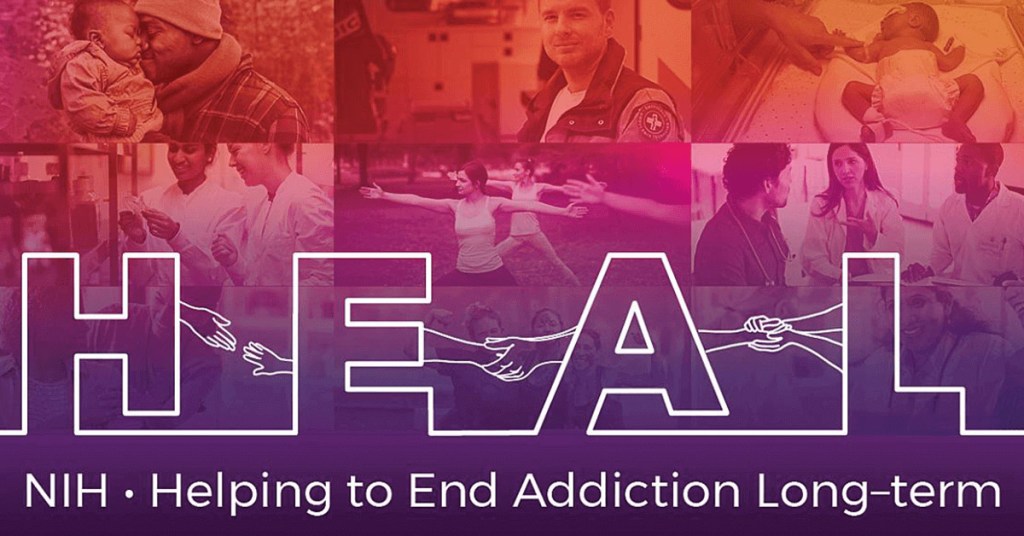
While the demands of the pandemic have been challenging for everyone, biomedical researchers have remained hard at work to address the opioid crisis. Among the many ways that NIH is supporting these efforts is through its Helping to End Addiction Long-Term (HEAL) Initiative, which is directing more than $1.5 billion to researchers and communities across the country.
Here’s a condensed transcript of our April 6th video dialogue, which focused on the impact of the COVID-19 pandemic on people struggling with substance use disorders and those who are trying to help them.
Collins: What have we learned so far through HEAL? Well, one thing HEAL is doing is tackling the need for pain treatments that help people avoid the risks of opioids. This research has uncovered new targets and therapeutics for different types of pain, including neuropathic, post-surgical, osteoarthritic, and chemotherapy induced. We’re testing implanted devices, such as electrodes and non-invasive nerve stimulation; and looking at complementary and integrative approaches, such as phone-based physical therapy for low back pain.
Through HEAL, we’ve launched a first-in-human test of a vaccine to protect against the harmful effects of opioids, including relapse and overdose. We’re also testing a tool that provides pharmacists with a validated opioid use disorder risk measure. The goal is to identify better who’s at high risk for opioid addiction and to determine what kind of early intervention could be put in place.
Despite COVID, many clinical studies are now recruiting participants. This includes family-based prevention programs, culturally tailored interventions for hard-hit American Indian populations, and interventions that address social inequities, such as lack of housing.
We are also making progress on the truly heart-breaking problem of babies born dependent on opioids. HEAL has launched a study to test the effectiveness of a new approach to care that measures the severity of a baby’s withdrawal, based on their ability to eat, sleep, and be consoled. This approach helps provide appropriate treatment for these infants, without the use of medication when possible. We’re also developing novel technologies to help treat neonatal opioid withdrawal syndrome, including a gently vibrating hospital bassinet pad that’s received breakthrough device designation from the FDA.
2020 was an extraordinary year that was tragic in so many ways, including lives lost and economic disasters that have fallen upon families. The resilience and ingenuity of the scientific community has been impressive. Quick pivoting has resulted in some gains through research, maybe you could even call them silver linings in the midst of this terrible storm.
Nora, what’s been at the forefront of your mind as we’ve watched things unfold?
Volkow: When we did this one year ago, we didn’t know what to expect. Obviously, we were concerned that the stressors associated with a pandemic, with unknowns, are factors that have been recognized for many years to increase drug use. Unfortunately, what we’ve seen is an increase in drug use of all types across the country.
We have seen an exacerbation of the opioid epidemic, as evidenced by the number of people who have died. Already, in the 12 months ending in July 2020, there was a 24 percent increase in mortality from overdoses. Within those numbers, there was close to a 50 percent increase in mortality associated with fentanyl. We’re also seeing an increase, not just in deaths from fentanyl and other synthetic opioids, but in deaths from stimulant drugs, like cocaine and methamphetamine. And the largest increases have been very much driven by drug combinations.
So, we have the perfect storm. We have people stressed to their limits by decreases in the economy, the loss of jobs, the death of loved ones. On the other hand, we see dealers taking the opportunity to bring in drugs such as synthetic opioids and synthetic stimulants and distribute them to a much wider extent than previously seen.

Collins: On top of that, people are at risk of getting sick from COVID-19. What have we learned about the risks of coronavirus illness for people who use drugs?
Volkow: It is a double whammy. When you look at the electronic health records about the outcomes of people diagnosed with substance use disorders, you consistently see an increased risk for getting infected with COVID-19. And if you look at those who get infected, you observe a significantly increased risk of dying from COVID.
What’s driving this vulnerability? One factor is the pharmacological effects of these drugs. Basically, all of the drugs of abuse that result in addiction, notably opioids, damage the cardiopulmonary system. Some also damage the immune system. And we know that individuals who have any disruption of cardiovascular health, pulmonary health, immune function, or metabolism are at higher risk of getting infected with COVID-19 and having adverse outcomes.
But there’s another factor that’s as important—one that’s very tractable. It is the way in which our society has dealt with substance use disorders: not actually treating them as a disease that requires intervention and support for recovery. The stigmatization of individuals with addiction, the lack of access to treatment, the social isolation, have all created havoc by making these individuals so much more vulnerable to get infected with COVID-19.
They will not go to a doctor. They don’t want to be stigmatized. They need to go out into the streets to get access to the drugs. Many times, they don’t have a choice of what drugs to take because they cannot afford anything except what’s offered to them. So, many, especially those who are minorities, end up homeless or in jails or prison. Even before COVID, we knew that prisons and jails are places where infections can transmit extraordinary rapidly. You could see this was going to result in very negative outcomes for this group of individuals.
Collins: Nora, tell us more about the trends contributing to the current crisis. Maybe three or four years ago, what was going straight up was opioid use, especially heroin. Then, fentanyl started coming up very fast and that has continued. Now, we are seeing more stimulants and mixing of different types of drugs. What is the basis for this?
Volkow: At the beginning of the opiate pandemic, mortality was mainly associated with white Americans, many in rural or semi-suburban areas of the Appalachian states and in New Mexico and Arizona. That has shifted. The highest increase in mortality from opioids, predominantly driven by fentanyl, is now among Black Americans. They’ve had very, very high rates of mortality during the COVID pandemic. And when you look at mortality from methamphetamine, it’s chilling to realize that the risk of dying from methamphetamine overdose is 12-fold higher among American Indians and Alaskan Natives than other groups. This should make us pause to think about what’s driving these terrible racial disparities.
As for drug combinations, many deaths from methamphetamine or cocaine—an estimated 50 percent—are linked to these stimulant drugs being combined with fentanyl or heroin. Dealers are lacing these non-opioid drugs with cheaper, yet potent, opioids to make a larger profit. Someone who’s addicted to a stimulant drug like cocaine or methamphetamine is not tolerant to opioids, which means they are going to be at high risk of overdose if they get a stimulant drug that’s laced with an opioid like fentanyl. That’s been contributing to the sharp rise in mortality from non-opioid drugs.
Collins: I’m glad you raised the issue of health disparities. 2020 will go down as a year in which our nation had to focus on three public health crises at once. The first is the crisis of opioid use disorder and rising mortality from use of other drugs. The second is COVID-19. And the third is the realization, although the problem has been there all along, that health disparities continue to shorten the lives of far too many people.
The latter crisis has little to do with biology, but everything to do with the way in which our society still is afflicted by structural racism. We at NIH are looking at this circumstance, realizing that our own health disparities research agenda needs to be rethought. We have not fully incorporated all the factors that play out in health inequities and racial inequities in our country.
You were also talking about how stimulants have become more widespread. What about treatments for people with stimulant use disorders?
Volkow: For opioid addiction, we’re lucky because we have very effective medications: methadone, buprenorphine, naltrexone. On top of that, we have naloxone, Narcan, that if administered on time, can save the life of a person who has overdosed.
We don’t have any FDA-approved medication for methamphetamine addiction, and we don’t have any overdose reversal for methamphetamine. At the beginning of this year, we funded a large clinical trial aimed at investigating the benefits of the combination of two medications that were already approved as anti-depressants and for the treatment of smoking cessation and alcoholism. It found this combination significantly inhibits the urge to take drugs and therefore helps people stay away from use of methamphetamine. Now, we want to replicate these findings, and to tie that replication study in with guidelines from the FDA on what is needed to approve our new indication for these medications. Why? Because then insurance can cover it, and that will increase the likelihood that people will get treated.
Another exciting possibility is a monoclonal antibody against methamphetamine that’s in Phase 2 clinical trials. If someone comes into the emergency room with an overdose of a combination of opioid and methamphetamine, naloxone often will not work. But this monoclonal antibody with naloxone may offer a greater likelihood of success.
Another thing that’s promising is that investigators have been able to modify monoclonal antibodies so they stay in the bloodstream for a longer time. That means we may someday be able to use this passive immunization approach as a treatment for methamphetamine addiction.
Collins: That’s good to hear. Speaking of progress, is there any you want to point to within HEAL?
Volkow: There’s a lot of excitement surrounding medication development. We’re interested in developing antidotes that will be more effective in reversing overdose deaths from fentanyl. We’re also interested in providing longer lasting medications for treatment of opioid use disorders, which would improve the likelihood of patients being protected from overdoses.
The Justice Community Opioid Innovation Network (JCOIN) is another HEAL landmark project. It involves a network of researchers that is working with judges and with the workers in jail and prison systems responsible for taking care of individuals with substance use disorders. Through this network, we’ve been able to start to harmonize practices. One thing that’s been transformative in the jail and prison system has been the embracing of telehealth. In the past, telehealth was not much of a reality in jails and prisons because of the fear of it could lead to communications that could perhaps be considered dangerous. That’s changed due to COVID-19. Now, telehealth is providing access to treatment for individuals in jail and prison, many of them with substance use disorders.
Also, because of COVID, many nonviolent individuals in jails and prisons were released. This gives us an opportunity to evaluate how best to help such individuals achieve recovery from substance use disorders. Hopefully we can generate data to show that there are much more effective strategies than incarceration for dealing with substance use disorders.
The HEALing Communities Study, involves Massachusetts, New York, Ohio, and Kentucky—four of the states with the highest rates of mortality from overdoses from the inception of the opioid epidemic. By implementing a battery of interventions for which there is evidence of benefit, this ambitious study set out to decrease overdose mortality by 40 percent in two years. Then, came COVID and turned everything upside down. Still, because we consolidated interactions between agencies, we’ve been able to apply support systems more efficiently in those communities in ways that have been very, very reinforcing. Obviously, there’ve been delays in implementation of interventions that require in-person interactions or that involve hospital emergency departments, which have been saturated with COVID patients.
We’ve learned a lot in the process. I may be too optimistic, but I do believe that we can stay on goal.
Collins: Now, I’d like to transition to a few questions from people who subscribe to the HEAL website. Announced at this meeting three years ago, the HEAL Initiative involves research participants and patients and stakeholders—especially people who have lived experience with pain, addiction, or both.
Let’s get to the first question: “What is NIH doing through HEAL to address the stigma that prevents people who need opioid medications for treatment from getting them?”
Volkow: A crucial question. As we look at the issue of stigma, we need to recognize that there are structural issues in how our society is prioritizing the importance of substance use disorders and the investments devoted to them. And we need to recognize that substance use disorder doesn’t exist in isolation; it is frequently comorbid with mental illness.
We need to listen. Some of the issues that we believe are most problematic are not. We need to empower these communities to speak up and help them do so. This is probably one of the most important things that we can do in terms of addressing stigma for addiction.
Collins: Absolutely. The HEAL Initiative has a number of projects that are focusing on stigma and coming up with tools to help reduce this. And here’s our second question: “In small communities, how can we provide more access to medications for opioid use disorder?”
Volkow: One project funded through HEAL was to evaluate the effectiveness of community pharmacies for delivering buprenorphine to individuals with opioid use disorder. The results show that patients receiving buprenorphine through community pharmacies in rural areas had as good outcomes as patients being treated by specialized clinicians on site.
Another change that’s made things easier is that in March 2020, the DEA relaxed its rules on how a physician can prescribe buprenorphine. In the past, you needed to go physically to see a doctor. Now, the DEA allows a patient to be initiated on buprenorphine through telehealth, and that’s opened the possibility of greater access to treatment in rural communities.
My perspective is let’s look at innovative ways of solving problems. Because the technology is changing in so many ways and so rapidly, let’s take advantage of it.
Collins: Totally with you on that. If there’s a silver lining to COVID-19, it’s that we’ve been forced to take stock of the ways we’ve been doing things. We will learn from this pandemic and change the way we approach so many things in health and medicine as a result. Certainly, opioid use disorder ought to be very high on that list. Let’s move on to another question: “What is the HEAL initiative doing to promote prevention of opioid use?”
Volkow: This is where the HEAL initiative is aiming to provide alternative treatments for the management of pain that reduce the risk of addiction.
Then there’s the issue of prevention in people who start to take opioids because they either want to get high or escape. With the COVID pandemic, we’ve seen increases in anxiety and in depression. Those are factors that can put a teenager or young adult on a trajectory for higher risk of substance use disorders.
So, what is HEAL doing? There is prevention research specifically targeted, for example, at the transition from adolescence to young adulthood. That is the period of greatest vulnerability of uptake of opioids, or drugs of misuse. We’re also targeting minority groups that may be at very, very high risk. We want to be able to understand the factors that make them more vulnerable to tailor prevention interventions more effectively.
Collins: Today, we’ve shared some of the issues that NIH is wrestling with in its efforts to address the crisis of opioid misuse and overdose, as well as other drugs that are now very much part of the challenge. To learn more, go to the HEAL website. You can also send us your thoughts through the HEAL Idea Exchange.
These developments give me hope in the wake of a very difficult year. Clearly, we still have the capacity to work together, we are resilient, and we are determined to put an end to our nation’s opioid crisis.
Volkow: Francis, I want to thank you for your incredible leadership and your support. I hope the COVID pandemic will bring forth a more equitable system, in which all people are given the chance for resilience that maximizes their life, happiness, and productivity. I think science is an extraordinary tool to help us do that.
Links:
Video: The 2021 Rx Drug Abuse & Heroin Summit: Francis Collins with Nora Volkow (NIH)
COVID-19 Research (NIH)
Helping to End Addiction Long-term (HEAL) Initiative (NIH)
HEAL Idea Exchange (NIH)
National Institute on Drug Abuse (NIH)
New Initiative Puts At-Home Testing to Work in the Fight Against COVID-19
Posted on by Dr. Francis Collins

Thankfully COVID-19 testing is now more widely available than it was earlier in the pandemic. But getting tested often still involves going to a doctor’s office or community testing site and waiting as long as a couple of days for the results. Testing would be so much easier if people could do it themselves at home. If the result came up positive, a person could immediately self-isolate, helping to stop the coronavirus that causes COVID-19, SARS-CoV-2, from spreading any further in their communities.
That’s why I’m happy to report that the Centers for Disease Control and Prevention (CDC), in close collaboration with state and local public health departments and with NIH, has begun an innovative community health initiative called “Say Yes! COVID Test.” The initiative, the first large-scale evaluation of community-wide, self-administered COVID-19 testing, was launched last week in Pitt County, NC, and will start soon in Chattanooga/Hamilton County, TN.
The initiative will provide as many as 160,000 residents in these two locales with free access to rapid COVID-19 home tests, supplied through NIH’s Rapid Acceleration of Diagnostics (RADx) initiative. Participants can administer these easy-to-use tests themselves up to three times a week for one month. The goal is to assess the benefits of self-administered COVID-19 testing and help guide other communities in implementing similar future programs to slow the spread of COVID-19.
The counties in North Carolina and Tennessee were selected based on several criteria. These included local infection rates; public availability of accurate COVID-19 tracking data, such as that gathered by wastewater surveillance; the presence of local infrastructure needed to support the project; and existing community relationships through RADx’s Underserved Populations (RADx-UP) program. Taken together, these criteria also help to ensure that vulnerable and underserved populations will benefit from the initiative.
The test is called the QuickVue At-Home COVID-19 Test. Developed with RADx support by San Diego-based diagnostic company Quidel, this test is easily performed with a nasal swab and offers results in just 10 minutes. Last week, the test was among several authorized by the Food and Drug Administration (FDA) for over-the-counter use to screen for COVID-19 at home.
Participants can order their QuickVue test kits online for home delivery or local pick up. A free online tool, which was developed with NIH support by CareEvolution, LLC, Ann Arbor, MI, will also be available to provide testing instructions, help in understanding test results, and text message reminders about testing. This innovative tool is also available as a smartphone app.
A recent study, supported by the RADx initiative, found that rapid antigen testing for COVID-19, when conducted at least three times per week, achieves a viral detection level on par with the gold standard of PCR-based COVID-19 testing processed in a lab [1]. That’s especially significant considering the other advantages of a low-cost, self-administered rapid test, including confidential results at home in minutes.
The Say Yes! COVID Test initiative is an important next step in informing the best testing strategies in communities all over the country to end this and future pandemics. The initiative will also help to determine how readily people accept such testing when it’s made available to them. If the foundational data looks promising, the hope is that rapid at-home tests will help to encourage people to protect themselves and others by following the three W’s (Wear a mask. Wash your hands. Watch your distance), getting vaccinated, and saying “Yes” to the COVID-19 test.
Reference:
[1] Longitudinal assessment of diagnostic test performance over the course of acute SARS-CoV-2 infection. Smith RL, Gibson LL, Martinez PP, Heetderks WJ, McManus DD, Brooke CB, et al. medRxiv, 2021 March 20.
Links:
CDC and NIH bring COVID-19 self-testing to residents in two locales, NIH News Release, March 31, 2021
Rapid Acceleration of Diagnostics (RADx) (NIH)
COVID-19 Testing (CDC)
Quidel Corporation (San Diego, CA)
Coronavirus (COVID-19) Update: FDA Continues to Advance Over-the Counter and Other Screening Test Development, FDA News Release, March 31, 2021
NIH Support: National Heart, Lung, and Blood Institute; National Institute of Biomedical Imaging and Bioengineering
Previous Page Next Page