Snapshots of Life: The Brain’s Microscopic Green Trash Bins
Posted on by Dr. Francis Collins
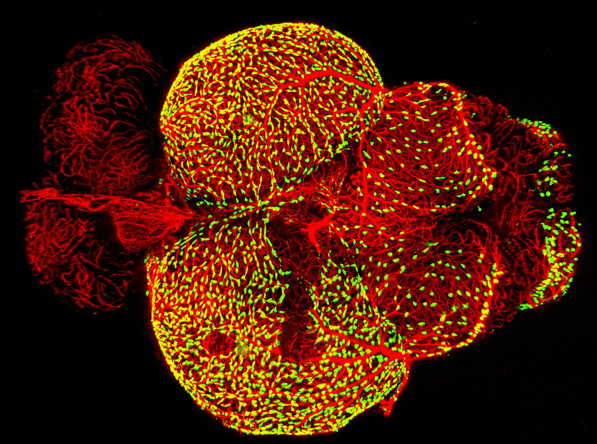
Credit: Marina Venero Galanternik, Daniel Castranova, Tuyet Nguyen, and Brant M. Weinstein, NICHD, NIH
There are trash bins in our homes, on our streets, and even as a popular icon on our desktop computers. And as this colorful image shows, trash bins of the cellular variety are also important in the brain.
This image—a winner in the Federation of American Societies for Experimental Biology’s 2017 BioArt competition—shows the brain of an adult zebrafish, a popular organism for studying how the brain works. It captures dense networks of blood vessels (red) lining the outer surface of the brain. Next to many of these vessels sit previously little-studied cells called fluorescent granular perithelial cells (yellowish green). Researchers now believe these cells, often shortened to FGPs, act much like trash receptacles that continuously take in and store waste products to keep the brain tidy and functioning well.
The image is the work of an NIH team including Marina Venero Galanternik and Daniel Castranova in Brant Weinstein’s laboratory at the Eunice Kennedy Shriver National Institute of Child Health and Human Development (NICHD), Bethesda, MD. The Weinstein lab studies how networks of blood and lymphatic vessels form during development.
The researchers had engineered the lymphatic vessels of transgenic zebrafish to express green fluorescent protein (GFP) to help visualize them. But after snapping some photos, Castranova noticed that, in addition to lymphatic vessels, some strange cells on the brain’s surface also expressed GFP. The researchers identified them as possibly being FGPs, which were then thought to be a type of immune-derived cell called a perivascular macrophage.
As published in the journal eLife, the NICHD team found that FGPs aren’t immune cells at all [1], but instead arise from primitive endothelial cells lining the inside of a specific set of blood vessels in the brain. After emerging from blood vessels, these cells migrate to the outermost layer of the brain, taking up residence next to superficial blood vessels. Once there, they appear to play an important role in ridding the brain of cellular waste products. Indeed, waste materials experimentally injected into the brain were quickly gobbled up by the FGPs.
While many questions remain, the researchers now suspect that in fish and mammals— including humans—these cells are an important part of the blood-brain barrier, the well-known functional deterrent that keeps drugs and other substances from traveling from the blood circulation into the functioning brain.In addition to sucking up cellular waste from inside the brain, FGPs may also take in viruses or toxins that might otherwise make their way from the bloodstream or the external environment into the brain. Now that the team has characterized these fascinating cells, including their origin in the blood vessels, they will learn more about their role in helping the brain cope with life’s many challenges—including infection, aging, and, of course, the need to take out the trash.
Reference:
[1] A novel perivascular cell population in the zebrafish brain. Venero Galanternik M, Castranova D, Gore AV, Blewett NH, Jung HM, Stratman AN, Kirby MR, Iben J, Miller MF, Kawakami K, Maraia RJ, Weinstein BM. Elife. 2017 Apr 11;6.
Links:
Video: Zebrafish Development (Eunice Kennedy Shriver National Institute of Child Health and Human Development/NIH)
Brant Weinstein (NIH/National Institute of Child Health and Human Development)
BioArt (Federation of American Societies for Experimental Biology, Bethesda, MD)
NIH Support: Eunice Kennedy Shriver National Institute of Child Health and Human Development; National Human Genome Research Institute
I have had a Grade 4 Glioblastoma diagnosis two and half years ago. Went through surgery to remove tumor, then 7 weeks of radiation while taking oral chemotherapy for the next 9 months. I have MRI’s every 2 to 3 months to look for any regrowth of tumor and I am still in the clear.
What, if any, does this study reflect on one with Glioblastoma ?