Snapshots of Life: Wired for Nerve Regeneration
Posted on by Dr. Francis Collins
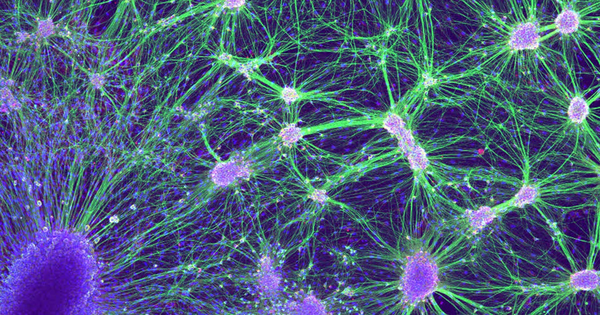
Credit: Laura Struzyna, Cullen Laboratory, Perelman School of Medicine, University of Pennsylvania, Philadelphia
Getting nerve cells to grow in the lab can be a challenge. But when it works, the result can be a thing of beauty for both science and art. What you see growing in the Petri dish shown above are nerve cells from an embryonic rat. On the bottom left is a dorsal root ganglion (dark purple), which is a cluster of sensory nerve bodies normally found just outside the spinal cord. To the right are the nuclei (light purple) and axons (green) of motor neurons, which are the nerve cells involved in forming key signaling networks.
Laura Struzyna, a graduate student in the lab of NIH grantee D. Kacy Cullen at the University of Pennsylvania’s Perelman School of Medicine, Philadelphia, is using laboratory-grown nerve cells in her efforts to learn how to bioengineer nerve grafts. The hope is this work will one day lead to grafts that can be used to treat people whose nerves have been damaged by car accidents or other traumatic injuries.
Here’s how the process currently works. After growing the cells in a Petri dish, Struzyna transfers them into a custom-built microdevice called a “mechanobioreactor.” There, the cells continue to grow for several days, while a small built-in motor applies controlled mechanical tension to the axons and progressively elongates them a few microns or more. (A micron is one millionth of a meter.) The goal is to prepare these cultured neurons for transplantation into an injured area, where their unusually long axons will stand out and attract nearby regenerating nerve cells.
Once a connection is made, the elongated axons serve as living scaffolds that provide the natural biological prompts needed to guide the regenerating nerve cells through the process of growing and linking to other cells to form a larger sensory network. If all goes well, the expanding network of new nerve fibers will fill in the injured area as new tissue forms and return sensation.
So far, things have gone beautifully in rat studies. For repairing nerve injuries measuring 1 to 5 centimeters in length, Struzyna says some of her bioengineered nerve grafts have performed as well as today’s gold-standard treatment, in which a patient’s own nerve tissue is transplanted into the affected area.
In addition to its encouraging implications for people suffering from nerve injuries, Struzyna’s work has also received artistic accolades. The image above, snapped with a confocal microscope, was among those recently featured in Perelman’s 2016 “Art in Science” competition.
Links:
Cullen Laboratory (Perelman School of Medicine, University of Pennsylvania, Philadelphia)
2016 Art in Science Competition Winners (Perelman School of Medicine)
NIH Support: National Institute of Neurological Disorders and Stroke
Such interesting things.
good article! thank you.