Snapshots of Life: Development in Exquisite Detail
Posted on by Dr. Francis Collins
Credit: Shachi Bhatt and Paul Trainor, Stowers Institute for Medical Research, Kansas City, MO
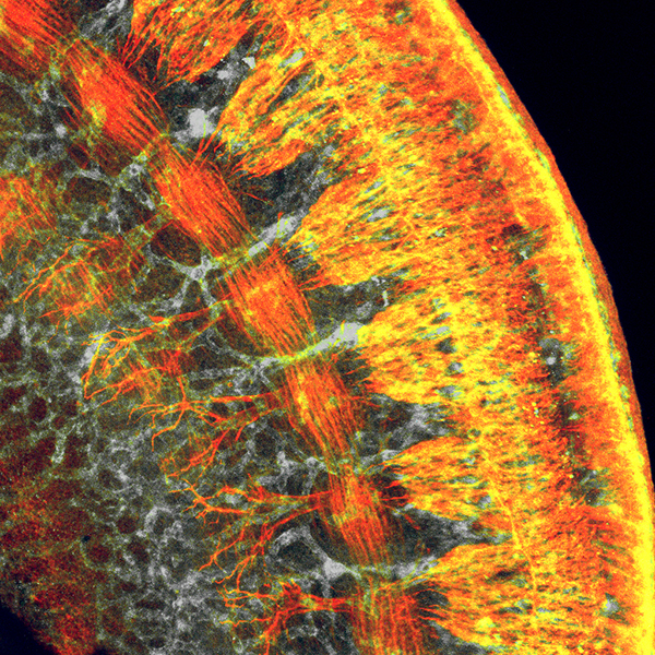
If you’ve ever tried to take photos of wiggly kids, you know that it usually takes several attempts before you get the perfect shot. It’s often the same for biomedical researchers when taking images with microscopes because there are so many variables—from sample preparation to instrument calibration—to take into account. Still, there are always exceptions where everything comes together just right, and you are looking at one of them! On her first try at using a confocal microscope to image this cross-section of a mouse embryo’s torso, postdoc Shachi Bhatt captured a gem of an image that sheds new light on mammalian development.
Bhatt, who works in the NIH-supported lab of Paul Trainor at the Stowers Institute for Medical Research, Kansas City, MO, produced this micrograph as part of a quest to understand the striking parallels seen between the development of the nervous system and the vascular system in mammals. Fluorescent markers were used to label proteins uniquely expressed in each type of tissue: reddish-orange delineates developing nerve cells; gray highlights developing blood vessels; and yellow shows where the nerve cells and blood vessels overlap.
While this award-winning image from the Federation of American Societies for Experimental Biology’s 2015 BioArt competition is focused on the torso, the Trainor lab’s main area of interest is development of the head and face. During their ongoing search for genes expressed in craniofacial development, Bhatt and her colleagues came across a gene that encodes a protein, called Med23, that plugs into a main developmental circuit that maintains needed patterns of gene expression. They went on to discover that Med23 not only is important in craniofacial development, it helps to guide the formation of both the nervous and vascular systems throughout the body.
While Bhatt continues to look into this unexpected link, her main research interest remains the possibility that Med23 might play a role in causing some cases of cleft lip and palate. Though this common birth defect usually is correctable with several surgeries, families undergo tremendous emotional and economic hardships during the process. Researchers are pushing ahead to understand the developmental complexity that underlies cleft lip and palate in hopes of learning how to prevent it. As Bhatt moves forward with this interesting lead, she’ll continue to take beautifully informative pictures.
Links:
Cleft Lip and Palate (National Institute of Dental and Craniofacial Research)
Paul Trainor (Stowers Institute for Medical Research, Kansas City, MO)
BioArt (Federation of American Societies for Experimental Biology, Bethesda, MD)
NIH Support: National Institute of Dental and Craniofacial Research
surely, you should be careful and attentive when working with these medical machines/instruments
I think cleft lip and palate is genetic.