Snapshots of Life: Portrait of Zika Virus
Posted on by Dr. Francis Collins
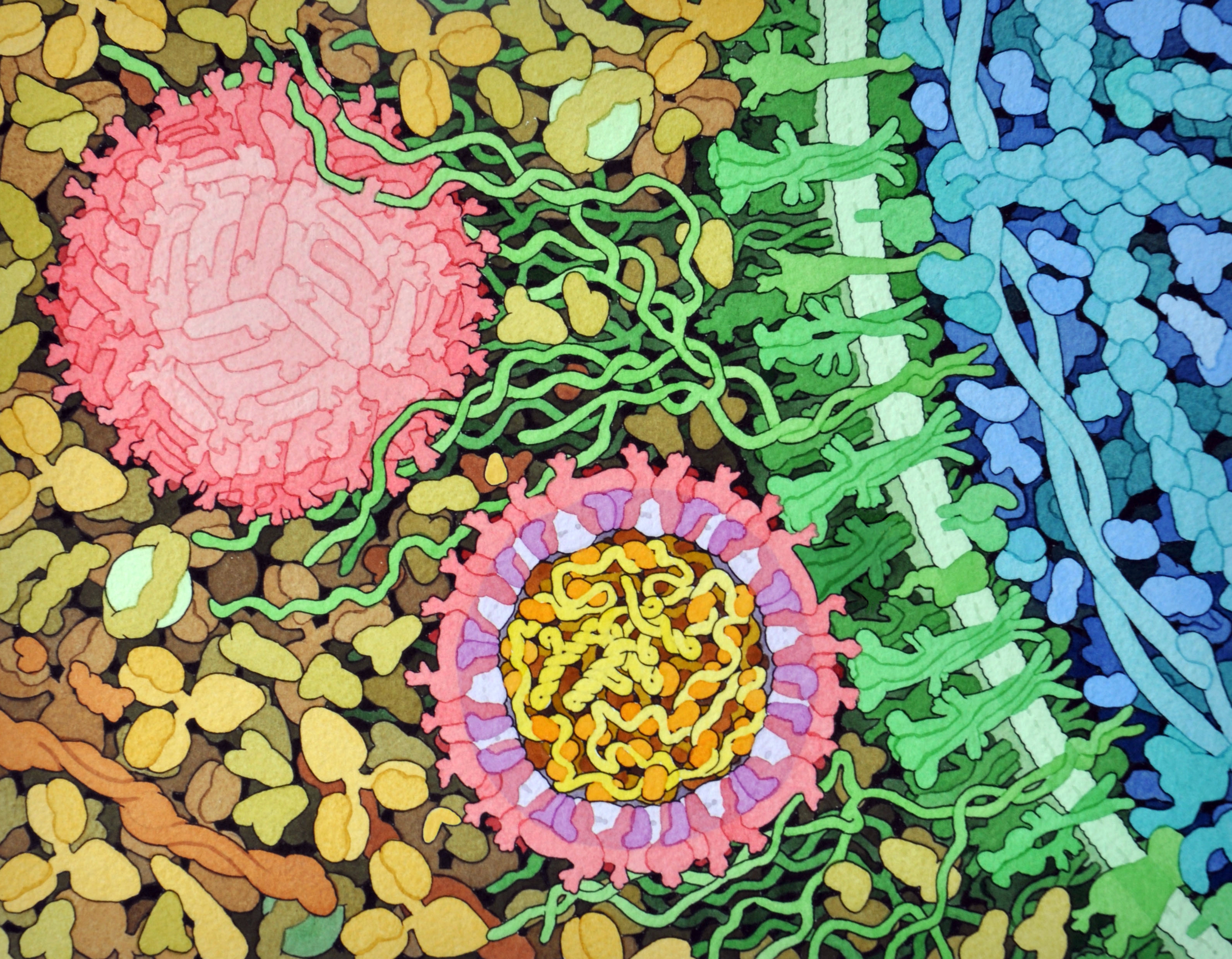
Credit: David Goodsell, The Scripps Research Institute
This lively interplay of shape and color is an artistic rendering of the Zika virus preparing to enter a cell (blue) by binding to its protein receptors (green). The spherical structures (pink) represent two Zika viruses in a blood vessel filled with blood plasma cells (tan). The virus in the middle in cross section shows viral envelope proteins (red) studding the outer surface, with membrane proteins (pink) embedded in a fatty layer of lipids (light purples). In the innermost circle, you can see the viral genome (yellow) coiled around capsid proteins (orange).
This image was sketched and hand-painted with watercolors by David Goodsell, a researcher and illustrator at The Scripps Research Institute, La Jolla, CA. Goodsell put paint and science to paper as part of the “Molecule of the Month” series run by RCSB Protein Data Bank (PDB), which NIH co-supports with the National Science Foundation and the Department of Energy. The PDB, which contains structural data on thousands of proteins and small molecules, uses its “Molecule of the Month” series to help students visualize a molecule or virus and to encourage their exploration of structural biology and its applications to medicine.
While the selection of Zika as May’s Molecule of the Month is surely related to its prominent place in world news, Goodsell drew heavily from the recently published near-atomic 3D map of the virus by a team of NIH grantees, led by Richard Kuhn’s group at Purdue University, West Lafayette, IN [1]. Solving the Zika structure marks an important step forward in efforts to develop a safe and effective vaccine against this emerging health threat.
In other news related to development of a Zika vaccine, Anthony Fauci, director of NIH’s National Institute of Allergy and Infectious Diseases (NIAID), recently announced plans to begin early human testing of an experimental vaccine later this year. If that vaccine proves safe and capable of spurring the desired immune response, Fauci indicated NIH would help to launch a larger clinical trial in a country with a very high rate of Zika infection early in 2017 [2].
That’s encouraging information for the hundreds of millions of people in our nation and around the world who are eagerly awaiting a Zika vaccine. In fact, a recent survey of more than 1,000 Americans conducted by the Annenberg Public Policy Center of the University of Pennsylvania, Philadelphia found that 55 percent of respondents would be “very” or “somewhat likely” to get a Zika vaccine—nearly as high as the percentage of people (62 percent) who got or intended to get a flu vaccine. While Zika infection during pregnancy has the potential to cause serious birth defects, the survey also found 45 percent of respondents were under the mistaken impression that someone who contracts Zika will likely die.
By the way, Goodsell’s scientific artistry goes back to the 1980s when researchers began solving the chemical structures of proteins at a faster pace. Goodsell wanted to capture the complexity of the molecular world. When he found the available computer programs inadequate, he pulled out his watercolors and started painting. More than 25 years later, Goodsell continues to earn high praise for his watercolor illustrations, while joining with his PDB colleagues to share them as an always-interesting Molecule of the Month.
References:
[1] The 3.8 Å resolution cryo-EM structure of Zika virus. Sirohi D, Chen Z, Sun L, Klose T, Pierson TC, Rossmann MG, Kuhn RJ. Science. 2016 Apr 22;352(6284):467-470.
[2] Zika vaccine efficacy trials could start in 2017. Cohen J. Science Insider, 2016 May 3.
Links:
Zika Virus (National Institute of Allergy and Infectious Diseases/NIH)
Richard J. Kuhn Lab (Purdue University, West Lafayette, IN)
David Goodsell (The Scripps Research Institute, La Jolla, CA)
Molecule of the Month, RCSB Protein Data Bank
Zika Virus (Annenberg Public Policy Center of the University of Pennsylvania, Philadelphia)
NIH Support: National Library of Medicine;National Institute of General Medical Sciences; National Cancer Institute
I’ve been out of the biology game for a while but back in the seventies it was believed that viruses were not cells but protein coating around DNA or RNA…The artistic rendering portrays the Zika viruses almost like cells with a lipid/protein membrane combination akin to the fluid-mosaic model for cell membranes with proteins floating in a lipid layer…Is the virus membrane now believed to be similar to a cell membrane?
Actually viruses are acellular ultra microorganisms, which are considered to be genetic material – either dna or rna, but never both are surrounded by protein coat called capsid. Viruses are obilgate parasites, which can multiply only for the host cell. Remember viruses don’t grow food or respire. Virus are simple structures that utilize ribosomes for the host cell enzyme and protein synthesis during reproduction. Viruses are specific to the host and its characteristics as living and non living thing … zika virus related as non envelop virus because lack of cell membrane. Remember virus only building block of cytoplasm.
Hi Hal–you’re correct on both counts–viruses are simpler than cells, since they can’t reproduce by themselves, and yes, some viruses have lipid membranes that are very similar to those that surround cells. The reason for this is that these types of viruses, termed “enveloped viruses”, bud from the surfaces of cells, taking a coat of membrane as they leave. Other enveloped viruses include influenza virus and HIV.
zika virus Thanks for the very useful and revealing article