Cool Videos: Reconstructing the Cerebral Cortex
Posted on by Dr. Francis Collins
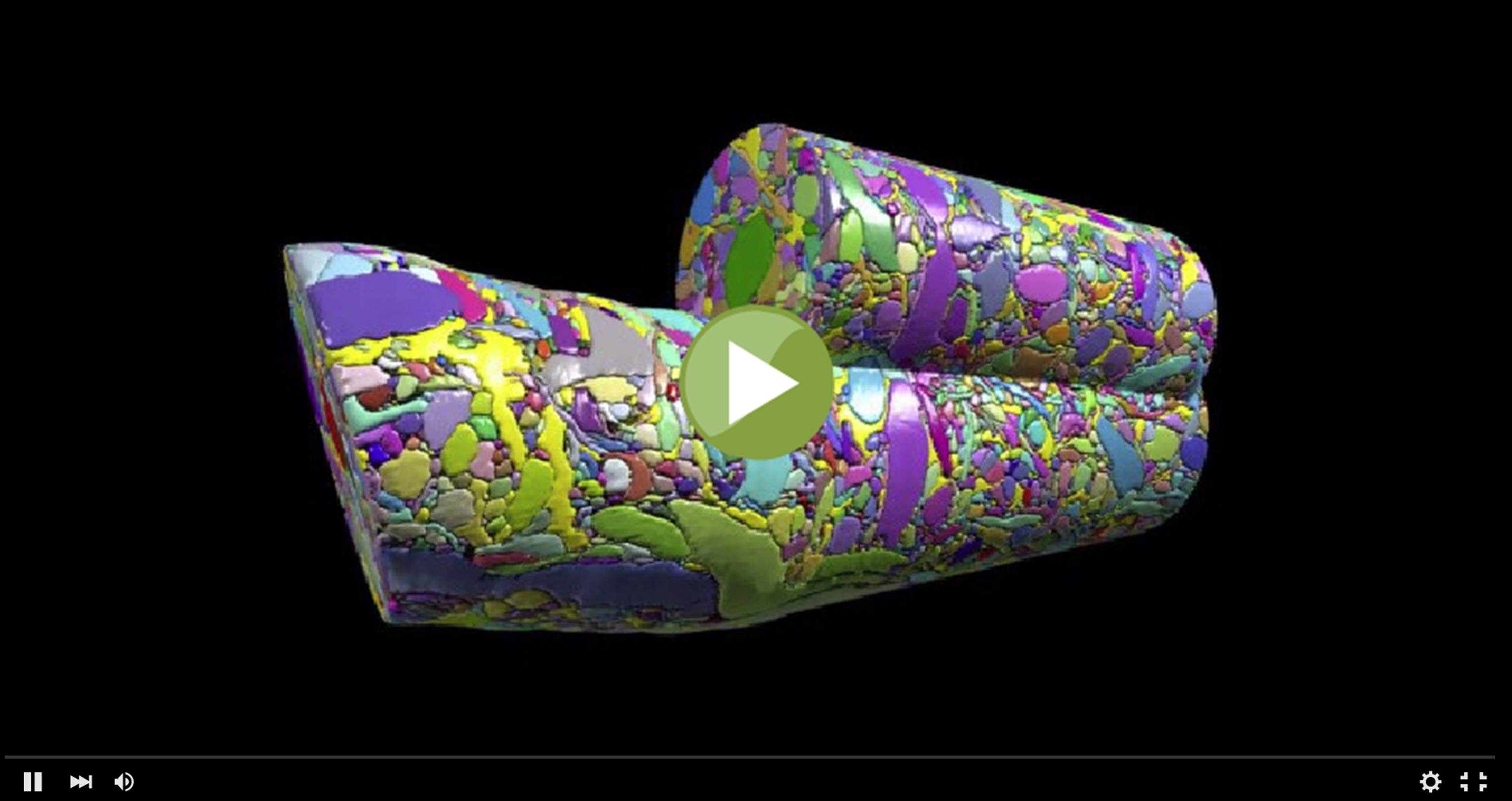
This colorful cylinder could pass for some sort of modern art sculpture, but it actually represents a sneak peak at some of the remarkable science that we can look forward to seeing from the Brain Research through Advancing Innovative Neurotechnologies (BRAIN) Initiative. In a recent study in the journal Cell [1], NIH grantee Jeff Lichtman of Harvard University, Cambridge, MA and his colleagues unveiled the first digitized reconstruction of tissue from the mammalian cerebral cortex—the outermost part of the brain, responsible for complex behaviors.
Specifically, Lichtman’s group mapped in exquisite detail a very small cube of a mouse’s cerebral cortex. In fact, the cube is so tiny (smaller than a grain of sand!) that it contained no whole cells, just a profoundly complex tangle of finger-like nerve cell extensions called axons and dendrites. And what you see in this video is just one cylindrical portion of that tissue sample, in which Licthtman and colleagues went full force to identify and label every single cellular and intracellular element. The message-sending axons are delineated in an array of pastel colors, while more vivid hues of red, green, and purple mark the message-receiving dendrites and bright yellow indicates the nerve-insulating glia. In total, the cylinder contains parts of about 600 axons, 40 different dendrites, and 500 synapses, where nerve impulses are transmitted between cells.
What’s fascinating is the researchers found that, contrary to what many have assumed, cortical nerve cells don’t appear to form connections based on proximity. Instead, the connections seem to be driven by cell identity, with certain axons preferring certain dendrites. Another intriguing finding was that once a good match is made, compatible axons and dendrites seem to forge not just one connection, but multiple connections.
So, how did these neuroscientists achieve their mapping feat? Among their many innovations was a creative device that resembles an old-fashioned movie projector. The gadget contains a diamond knife that slices a sample of brain tissue into extremely thin (29 nanometer) sections. After the sections are cut, they drop into a dish of water, where they bob to the surface and eventually attach to a continuously moving reel of tape. The researchers then cut the reel of tape into 2,250 individual strips of tissue, mounted them on silicon wafers, and moved them onto a scanning electron microscope to be photographed at extremely high resolution.
Other innovations came on the computational front. Daniel Berger and other team members devised software to digitize the images, align them properly, and recapitulate the natural 3D structure of the tissue. To help with this extremely difficult task, Berger created a computer program to segment elements in each tissue slice and outline them, much as images are outlined in a kid’s coloring book. This enabled researchers to “color in” each element in a distinctive hue that allowed them to differentiate one type of a dendrite from another, a dendrite from an axon, and so on.
But computers couldn’t do everything—some tasks required a human eye. For example, the researchers drew on the help of a number of student interns to analyze the high-resolution electron micrographs. One of their jobs: count the scatter of tiny membrane enclosed spheres at the end of each synapse. These spheres (which aren’t visible in this particular video—just one of 14 movies featured in the Cell paper) are vesicles containing the neurotransmitters that nerve cells use to communicate.
In total, Lichtman and colleagues mapped 1,700 synapses in their tiny cube of mammalian cortex. Those data have been made publicly available to researchers for further study via the Open Connectome Project, which is partially supported by NIH.
As remarkable as this work is, it also underscores how enormously challenging it will be to map the entire human brain. The tissue that was reconstructed in this study represents just 0.000001 percent of the mouse brain, and, even with a large and highly skilled team, it took about six years to map. Still, this work provides an important proof of principle that with sufficient resources, technological innovation, and scientific ingenuity, the BRAIN Initiative can attain its ambitious goal of producing a dynamic picture of the human brain that, for the first time, shows how individual cells and complex neural circuits interact in both time and space.
Reference:
[1] Saturated Reconstruction of a Volume of Neocortex. Kasthuri N, Hayworth KJ, Berger DR, Schalek RL, Conchello JA, Knowles-Barley S, Lee D, Vázquez-Reina A, Kaynig V, Jones TR, Roberts M, Morgan JL, Tapia JC, Seung HS, Roncal WG, Vogelstein JT, Burns R, Sussman DL, Priebe CE, Pfister H, Lichtman JW et al. Cell 2015 Jul 30;162(3):648-661.
Links:
Lichtman Lab (Harvard University, Cambridge, MA)
A boost for understanding the brain: Harvard researchers among the first to receive grant funding through BRAIN Initiative. Peter Ruell. Harvard Gazette. 30 September 2014.
Video: Connectomics (Jeff Lichtman, TEDx Talk, January 18, 2013)
The BRAIN Initiative (NIH)
Open Connectome Project (Johns Hopkins University, Baltimore)
The Human Connectome Project (NIH Blueprint for Neuroscience Research)
NIH Support: National Institute of Neurological Disorders and Stroke; National Institute of Mental Health; Common Fund
Litchman’s research studies are truly astounding. Eagerly waiting to see the fruits of his research helping the brain damaged patients.